. 2001 Aug 15;21(16):6264-73.
doi: 10.1523/JNEUROSCI.21-16-06264.2001.

Increasing the number of synapses modifies olfactory perception in Drosophila

Affiliations

Increasing the number of synapses modifies olfactory perception in Drosophila

A Acebes et al. J Neurosci. .

Abstract

The Drosophila mutant gigas produces an enlargement of postmitotic cells caused by additional rounds of DNA replication. In neurons, the mutant cell establishes more synapses than normal. We have taken advantage of this feature to study the effect of synapse number on odorant perception. Mosaic adults were generated in which one antenna was homozygous for gigas, whereas the contralateral side served as an internal control. Morphological analysis indicates that the number and type of sensory afferents forming the mutant antenna, as well as their projection to the olfactory glomeruli, are normal. In contrast, the volume of identified glomeruli increases to a variable extent, and mutant sensory neurons branch profusely. The number of synapses, estimated in the ventral (V) glomerulus that receives ipsilateral afferents only, is increased twofold to threefold. Large-dense-core vesicle-containing terminals that probably modulate olfactory centers are identified in the V glomerulus. Their number and size are not modified by the mutant input. Sensory transduction, measured by electroantennograms, is normal in amplitude and kinetics. In odorant tests, however, the profile of the behavioral response to ethyl acetate shows attractive responses to concentrations to which sibling controls remain indifferent (10(-)8 and 10(-)7 v/v). In addition, the intensity of the response is augmented both at attractive and repulsive odorant concentrations with respect to that of controls. These results demonstrate that increased synapse number in the sensory neurons can modify the behavior of the organism, allowing a higher sensitivity of perception.


Figures
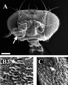
Fig. 1.
Fig. 1.
Morphology of antennal gig mosaics. Scanning electron microscope images from aM+gig antennal clone.A, Frontal view of a mosaic head showing the size difference between the gig (arrow) and contralateral antennae. B, gig sensillae are also larger than those from the contralateral side (C) (arrows indicate trichoid sensillae). Scale bar: A, 100 μm; B,C, 10 μm.
Fig. 2.
Fig. 2.
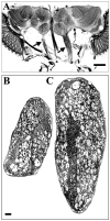
Antennal nerves of gig mosaics.A, Horizontal section from a gig mosaic head stained by Holmes-Blest silver impregnation. Note the diameter difference between normal (left) andgigas (right) nerves (arrows). See also Figure 3B. Anterior is down. B, C, Ultrathin frontal sections from normal (B) and mutant (C) antennal nerves from the same mosaic. The plane of section is approximately at the level of arrowsin A. Note the caliber of gig neurons, approximately twice that of its contralateral homologs. Both nerves are flattened from the lateral sides, but the number of axons is not significantly different (Table 1). Dorsal is up in B andC. Scale bars: A, 50 μm;B, C, 2 μm.
Fig. 3.
Fig. 3.
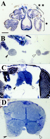
Brain structural effects of an antennal mosaic. Toluidine-stained semithin (1 μm) frontal sections through a single mosaic. A, Section at the level of antennal segments showing the funiculus (asterisk) and the pedicel (double asterisk). The mutant clone includes the whole antenna on the left side. B, Section at 3 μm from that shown in A, showing the antennal nerves in the mutant (solid arrowhead) and normal (open arrowhead) sides.C, Section at 5 μm from A, showing the antennal nerves and lobes (stippled in the mutant side).D, Section at 9 μm from A. Note the larger area of the antennal nerve and lobe in the mutant side. Both lobes are enlarged with respect to the wild type because of the bilateral projection of a fraction of sensory afferents. The increment, however, is larger in the lobe ipsilateral to the mutant antenna because not all afferents have bilateral projections, and branching is usually more abundant in the ipsilateral glomeruli. This differential branching is present in wild type and maintained in the mutant (Fig.4). Scale bar: A, C, 50 μm;B, D, 25 μm.
Fig. 4.
Fig. 4.
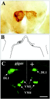
Projection pattern into antennal lobes.A, Frontal view of a whole mount C-S wild-type brain backfilled with HRP from the antenna on the left side. Note that most sensory afferents project and branch more intensively into the ipsilateral lobe. The V glomerulus is located in the ventral (down) apex of the lobe. Note that the contralateral locus is not marked by HRP. B, Camera lucida drawing of a gigas sensory neuron impregnated by the Golgi method. The mutant cell branches more profusely in the glomerulus ipsilateral to the mutant antenna (left side) as do normal cells. The amount of branching, however, is higher than in the wild type (Stocker et al., 1990). C, Frontal view of a confocal microscope section from the brain of a mosaic induced in a Gal4–72OK/UNG6 background. This Gal4 drives the expression of ann-Syn-GFP chimera in olfactory neurons that project to DL1, VM1, and VM4 glomeruli (arrows). The mutant afferents maintain this projection pattern. Note, however, the increased size of glomeruli ipsilateral to the mutant antenna. Lobes ipsilateral to mutant (gigas) and normal (+) antennae are separated by a dotted line. Scale bar:A, 65 μm; B, 40 μm; C, 30 μm.
Fig. 5.
Fig. 5.
Synaptic contacts in the olfactory neuropil. Electron micrograph of a V glomerulus innervated from agig antenna. Several presynaptic active zones are marked by framed arrowheads. Their size is normal, but their number is increased (Table 2). Scale bar, 500 nm.
Fig. 6.
Fig. 6.
Olfactory transduction tests. A,EAGs obtained in response to five ethyl acetate concentrations (105 to 101) from sibling controls (n = 6), and gig mosaics (n = 5). Amplitudes correlate with stimulus concentrations. B, Logarithmic plot of EAG amplitudes from sibling controls (filled inverted triangles), contralateral (filled circles), and gig mosaics (open squares). No significant difference is observed between mutant and controls. Error bars indicate SEM.
Fig. 7.
Fig. 7.
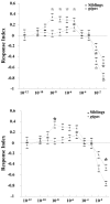
Olfactory behavioral responses. Dose–response curves from experimental mosaics (gray squares) and sibling controls (black diamonds). Values represent the mean response (±SEM) from 70 individuals per odorant concentration. Olfactory index values can range from 1 (full attraction) to −1 (full repulsion), and 0 is considered the indifference line.A, Olfactory responses significantly different (χ2 test) from indifference. These were found at 101 and 102 in sibling controls (p < 0.05) and mosaics (p < 0.01), and at 108 to 105 in the mosaics (p < 0.05, p < 0.01,p < 0.05, p < 0.05, respectively). Note the significant attraction response of mosaics at concentrations that yield indifference in the controls (108 to 105). B,Significant differences (Mann–Whitney U test) between mosaics and sibling controls at 108 and 101 concentrations (p < 0.05 and p < 0.01, respectively). Note the augmented responses in the mutant.

References

    1. Alcorta E. Characterization of the electroantennogram in Drosophila melanogaster and its use for identifying olfactory capture and transduction mutants. J Neurophysiol. 1991;65:702–714. - PubMed
    1. Anton S, Homberg U. Antennal lobe structure. In: Hansson BS, editor. Insect olfaction. Springer; Berlin: 1999. pp. 98–125.
    1. Ayer RK, Jr, Carlson J. Olfactory physiology in the Drosophila antenna and maxillary palp: acj6 distinguishes two classes of odorant pathways. J Neurobiol. 1992;23:965–982. - PubMed
    1. Ayyub C, Paranjape J, Rodrigues V, Siddiqi O. Genetics of olfactory behavior in Drosophila melanogaster. J Neurogenet. 1990;6:243–262. - PubMed
    1. Bailey CH, Kandel ER. Structural changes accompanying memory storage. Annu Rev Neurosci. 1993;55:397–426. - PubMed

Publication types

MeSH terms