. 2001 Aug 15;21(16):6308-20.
doi: 10.1523/JNEUROSCI.21-16-06308.2001.

Adenosine A1 receptors reduce release from excitatory but not inhibitory synaptic inputs onto lateral horn neurons

Affiliations

Adenosine A1 receptors reduce release from excitatory but not inhibitory synaptic inputs onto lateral horn neurons

S A Deuchars et al. J Neurosci. .

Abstract

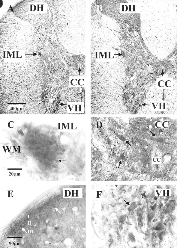
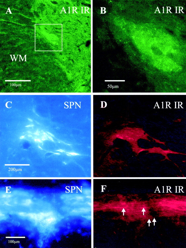
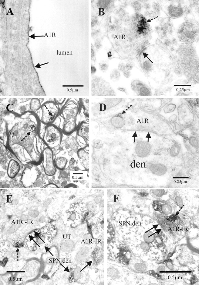
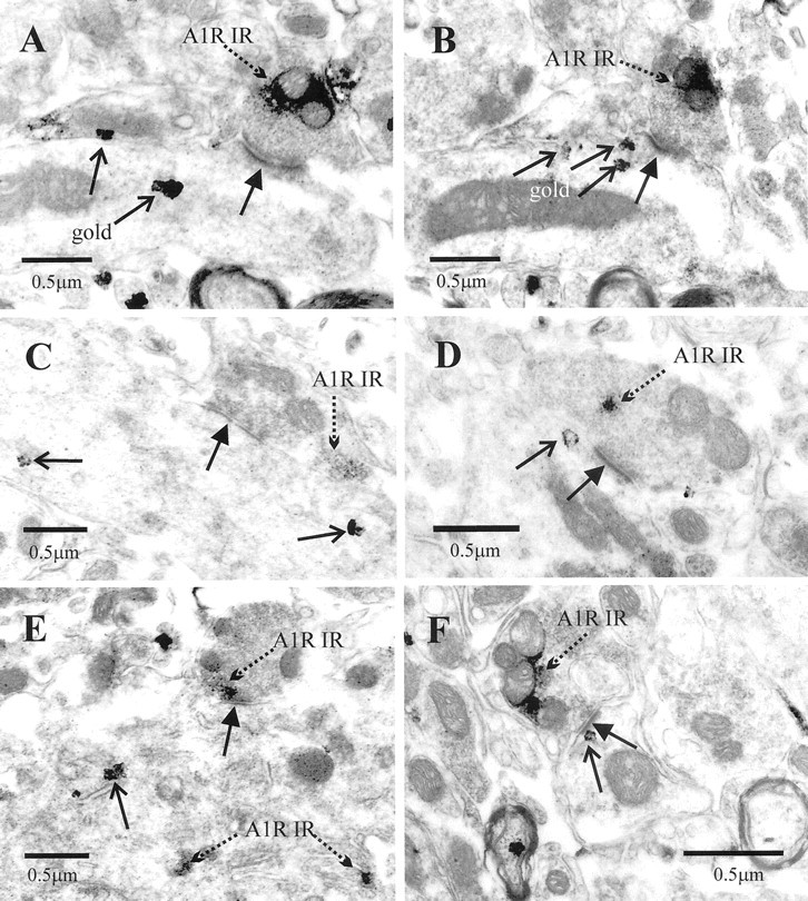
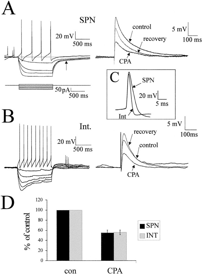
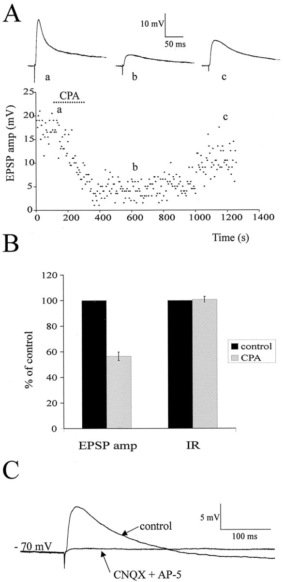
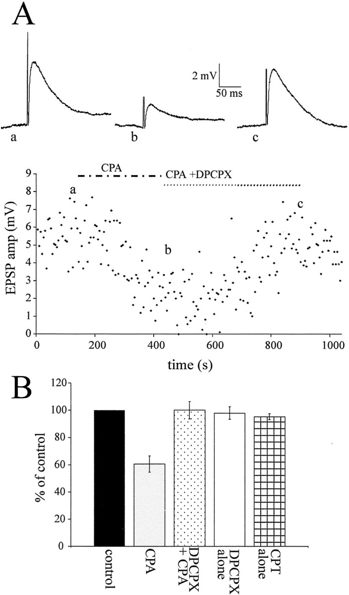
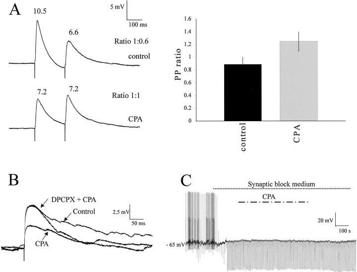
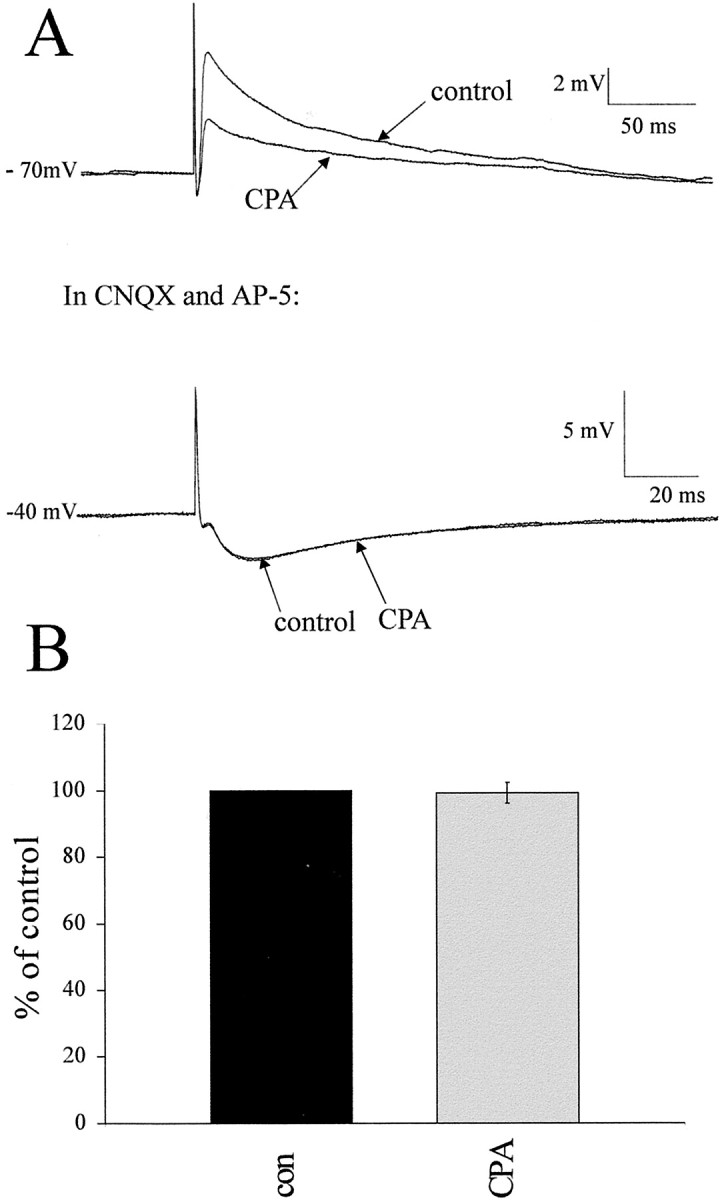
Although adenosine is an important neuromodulator in the CNS, its role in modulating sympathetic outflow at the level of the spinal cord has not been studied. Because very little is known about adenosine A1 receptors (A1Rs) in the spinal cord, we determined their location and role with particular reference to the control of sympathetic preganglionic activity and interneuronal activity in the rat. High levels of immunoreactivity for A1Rs were observed throughout the spinal cord. Immunostaining was dense in the intermediolateral cell column (IML) and intercalated nucleus, regions containing retrogradely labeled sympathetic preganglionic neurons (SPNs). Electron microscopy revealed A1R immunoreactivity (A1R-IR) within presynaptic terminals and (to a lesser extent) postsynaptic structures in the IML, as well as the luminal membrane of endothelial cells lining capillaries. Using double-labeling techniques, some presynaptic terminals were observed to synapse onto SPNs. To investigate the effects of activating these A1Rs, visualized whole-cell patch-clamp recordings were made from electrophysiologically and morphologically identified SPNs and interneurons. Applications of the A1R agonist cyclopentyladenosine (CPA) reduced the amplitude of EPSPs elicited by stimulation of the lateral funiculus, an effect blocked by the A1R antagonist 8-cyclopentyl-1,3-dipropylxanthine. These effects were attributable to adenosine acting at a presynaptic site because CPA application increased the paired-pulse ratio. CPA did not affect evoked IPSPs. These data show that activating A1Rs reduces fast excitatory, but not inhibitory, transmission onto SPNs and interneurons in the IML and that A1Rs may play a protective role on neurons involved in the control of sympathetic outflow.


Figures

Fig. 1.
Fig. 1.
Adenosine A1 receptor immunoreactivity in the thoracic spinal cord. A, Low magnification of staining obtained with the A1 receptor antibody raised in rabbit (gift from Dr. Mike Yates, Leeds University) in the thoracic spinal cord. Immunoreactivity was visualized with diaminobenzidine. Labeling was observed throughout the spinal cord. Even at low magnification, the IML is clearly seen as heavily stained. Staining is also dense around the central canal (CC), the dorsal horn (DH), and the ventral horn (VH). B, Low magnification of staining obtained with the A1 receptor antibody raised in goat (Santa Cruz Biotechnology), prepared identically to the material inA, except for the primary antibody. Note that the pattern of staining is almost identical, with the IML and ventral horn being particularly prominent. C, High magnification of the IML. Staining was a compact collection of punctate structures covering the IML adjacent to the white matter (WM). The arrow indicates one such punctate structure. D, In the vicinity of the central canal, labeling could be observed in the somata and dendrites of neurons (arrows), as well as presumptive fibers.E, In the dorsal horn, staining of fibers was dense in lamina II, and labeled neuronal somata could also be observed in laminas II and III (arrows). F, In the ventral horn, labeled fibers, somata, and dendrites of large neurons (arrow) were observed.
Fig. 2.
Fig. 2.
Fluorescence images indicating that adenosine A1 receptor immunoreactivity in the IML is dense in the region of retrogradely labeled SPNs. A, Low magnification of adenosine A1R-IR detected with the antibody raised in goat and visualized with Alexa488 viewed through a FITC filter set. The IML contains dense labeling and so stands out from surrounding structures. The boxed area surrounding the IML is shown at higher magnification in B.B, Higher magnification of the boxed areain A. At this magnification, it is clear that the staining in the IML has a punctate appearance. C, SPNs retrogradely labeled in the IML by an intraperitoneal injection of Fluorogold and visualized by UV illumination in a coronal section. SPNs appear blue–white under these conditions and contain Fluorogold in cell bodies and dendrites. The same section is shown inD. D, A1R-IR in the same section asC, detected with Cy3-conjugated secondary antibodies and viewed through a Cy3 filter set so that immunoreactivity for the adenosine A1R appears red. The A1R-IR makes the IML stand out as brighter red than the surrounding neuropil, and this bright staining persists into the lateral funiculus.E, SPNs retrogradely labeled in the IML by an intraperitoneal injection of Fluorogold and visualized by UV illumination in a longitudinal section. The same section is shown inF. F, A1R-IR in the same area of the section as E but viewed through a Cy3 filter set so that immunoreactivity for the adenosine A1 receptor appearsred. The A1 immunoreactivity runs in fibers along the IML (arrows).
Fig. 3.
Fig. 3.
Electron microscopic localization of the adenosine A1R-IR in the IML. A, A capillary in which A1R-IR is highly targeted to the luminal membrane of endothelial cells of blood vessels (arrows). B, A presynaptic terminal in the IML containing immunoreactivity for the A1R adjacent to the membrane (broken arrow indicates immunoreaction product). This terminal forms an asymmetric-type synaptic contact (arrow) with a dendritic structure.C, A1R-IR (broken arrows) was detected in numerous myelinated fibers in the lateral funiculus. D, A presynaptic terminal in the IML containing A1R-IR adjacent to the membrane (broken arrow indicates immunoreaction product). This terminal forms an asymmetric-type synaptic contact (arrows) with a dendritic structure (den). E, F, A1R-IR terminals formed synaptic contacts (arrows) with structures identified as SPN dendrites (SPN den) by the presence of crystalline reaction product (arrows) as a result of retrograde labeling with cholera toxin B chain, which was visualized with the TMB method. Note that the A1R-IR (broken arrows) is adjacent to the plasma membrane but some distance from the synaptic face.UT, unlabeled terminal.
Fig. 4.
Fig. 4.
Adenosine A1R-IR terminals form synaptic contacts with structures identified as SPNs by retrograde labeling.A, An A1R-IR terminal (broken arrow) forms an asymmetric-type synaptic contact (arrow) with a dendritic structure that contains silver-intensified gold particles (open arrows), indicating that it is a retrogradely labeled structure. The same terminal is shown inB. Note that the reaction product is adjacent to the membrane of the terminal but some distance from the active zone.B, The same terminal shown in A but serially several sections on, confirming that the dendrite is retrogradely labeled. The section has been tilted slightly with the goniometer to visualize the synaptic specialization. The silver grains in this section (open arrows) are spatially separate to those in A, indicating that these are different particles and not the result of intensification of the initial gold particles throughout the dendrite. C, D,F, Synaptic terminals (A1R-IR) containing A1 receptor immunoreactivity form synaptic contacts (arrows) with dendrites identified as retrogradely labeled SPNs by the silver-intensified gold particles (open arrows). Immunoreactivity is both adjacent to the membrane of labeled terminals (C, F) and within the cytoplasm (D). E, An A1R-IR terminal forms a synaptic contact (arrow) with the soma of an SPN. The soma contains not only silver-intensified gold particles (open arrows) but also A1R-IR associated with the endoplasmic reticulum (broken arrow).
Fig. 5.
Fig. 5.
CPA reduces the amplitude of EPSPs elicited in both SPNs and interneurons. A, On the leftare the voltage responses of an SPN to hyperpolarizing (averages of 3 sweeps) and depolarizing (single sweep) current pulses. Thetraces show a delayed return to resting potential at the end of the hyperpolarizing current pulses, indicative of activation of an IA (arrow). The action potential duration was 7.2 msec, and the afterhyperpolarization was quite simple with two components. On the right are average traces (of 10 sweeps) of the EPSP elicited by lf stimulation in control solution, CPA (100 nm), and then after switching back to standard aCSF. The EPSP amplitude was decreased by CPA application. B, The left shows the voltage responses of an interneuron to the same hyperpolarizing (averages of 3 sweeps) and depolarizing (single sweep) current pulses. At hyperpolarized potentials, a sag in the voltage response was observed, suggesting that an IH was activated. The action potential duration was 3.2 msec, and the AHP shows a distinct fast and slower phase. On the right are average traces (of 10 sweeps) of the EPSP elicited in the interneuron by lf stimulation. The EPSP amplitudes in control aCSF are not significantly different from those elicited in the SPN. In addition, the effects of CPA on the interneuronal EPSP are similar to those observed in the SPN. C, Comparisons of the duration of the interneuronal and SPN action potentials. D, Pooled data showing that the effects of CPA on the EPSPs elicited in SPNs are not significantly different from those in interneurons.
Fig. 6.
Fig. 6.
Time course of action of CPA and pharmacology of the EPSP. A, Graph of the amplitude of the EPSP elicited by lf stimulation in an SPN over time with sample EPSPs shown at thetop for each of the conditions (average of 10 consecutive sweeps). It can be clearly seen that CPA application caused a decrease in EPSP amplitude that slowly recovered as the CPA was washed off. B, Pooled data from 26 neurons showing the effect of CPA on EPSP amplitude and input resistance. CPA caused a significant decrease in EPSP amplitude but had no significant effect on input resistance. C, In another SPN, the EPSP was abolished by application of the excitatory amino acid receptor antagonists CNQX (20 μm) and AP-5 (50 μm), indicating that the EPSP is mediated by activation of these receptors postsynaptically.
Fig. 7.
Fig. 7.
DPCPX antagonized the effects of CPA.A, Graph of the EPSP amplitude elicited by lf stimulation against time in an interneuron with traces of the EPSP above (averages of 10 sweeps). CPA (100 nm) decreased the EPSP amplitude, an effect that was antagonized by application of DPCPX (300 nm), the A1R antagonist, together with CPA.B, Pooled data showing the effect of CPA on the EPSP and the antagonism by DPCPX, which restored the EPSP to the control amplitude. Both DPCPX and CPT (10 μm), another A1R antagonist, had no effect on EPSP amplitude when applied alone.
Fig. 8.
Fig. 8.
Effects of CPA are attributable to a presynaptic action. A, Responses of an SPN to twin pulse stimulation of the lf (200 msec apart) in control solution showed paired-pulse depression (i.e., the response to the second pulse was smaller than the first). In CPA, the second response is the same size as the first reduced response, with the ratio now being 1:1. On theright are the pooled data showing the paired-pulse ratio increases in the presence of CPA, indicative of a presynaptic site of action for adenosine. B, Effects of applications of CPA and DPCPX plus CPA in a neuron recorded using a patch solution in which cesium sulfate is the main component. CPA decreased the EPSP amplitude without effect on the membrane potential. This reduction was antagonized by DPCPX. C, In synaptic block medium containing TTX, DNQX, AP-5, strychnine, and bicuculline, action potentials and the response to lf stimulation were blocked. Hyperpolarizing current pulses were then applied to check input resistance, and CPA was applied. There was no change in membrane potential or input resistance with application of CPA in this recording medium.
Fig. 9.
Fig. 9.
Fast IPSPs elicited by lf stimulation are not affected by CPA. A, Reponses of an interneuron to lf stimulation in control and CPA-containing aCSF. CPA significantly reduced the EPSP amplitude. The EPSP was then antagonized by application of CNQX and AP-5 to reveal an underlying IPSP (seen here at −40 mV), which increased in amplitude as the membrane was depolarized. This IPSP was not affected by CPA application at the same concentration (100 nm) as that which reduced the EPSP. B, Pooled data from five neurons in which an IPSP was revealed. In all five cases, the IPSP was not affected by CPA, suggesting a selective site of action of the adenosine.

References

    1. Ackley MA, Baldwin SA, King AE. Inhibition of adenosine uptake attenuates glutamatergic synaptic transmission in lamina II of the rat spinal cord in vitro. Soc Neurosci Abstr. 2000;26:821.5.
    1. Alkadhi KA, Brown TR, Sabouni MH. Inhibitory effect of adenosine on transmission in sympathetic ganglia. Naunyn Schmiedebergs Arch Pharmacol. 1984;328:16–19. - PubMed
    1. Bagley EE, Vaughan CW, Christie MJ. Inhibition by adenosine receptor agonists of synaptic transmission in rat periaqueductal grey neurons. J Physiol (Lond) 1999;516:219–225. - PMC - PubMed
    1. Chau D, Johns DG, Schramm LP. Ongoing and stimulus-evoked activity of sympathetically correlated neurons in the intermediate zone and dorsal horn of acutely spinalized rats. J Neurophysiol. 2000;83:2699–2707. - PubMed
    1. Choca JI, Proudfit HK, Green RD. Identification of A1 and A2 adenosine receptors in the rat spinal cord. J Pharmacol Exp Ther. 1987;242:905–910. - PubMed

Publication types

MeSH terms

LinkOut - more resources