. 2001 Aug 15;21(16):6413-22.
doi: 10.1523/JNEUROSCI.21-16-06413.2001.

The contribution of activity-dependent synaptic plasticity to classical conditioning in Aplysia

Affiliations

The contribution of activity-dependent synaptic plasticity to classical conditioning in Aplysia

I Antonov et al. J Neurosci. .

Abstract

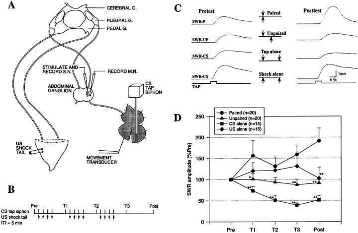
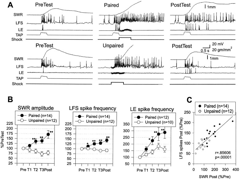
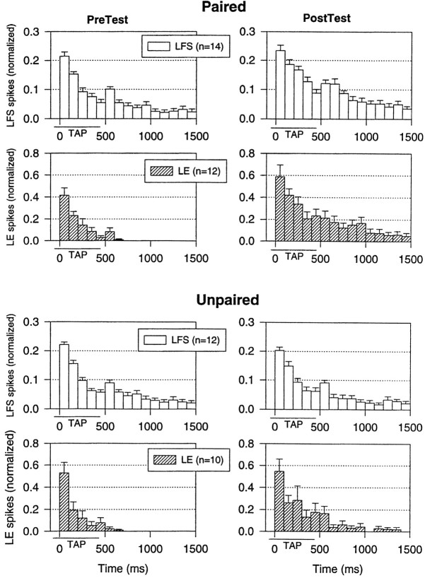
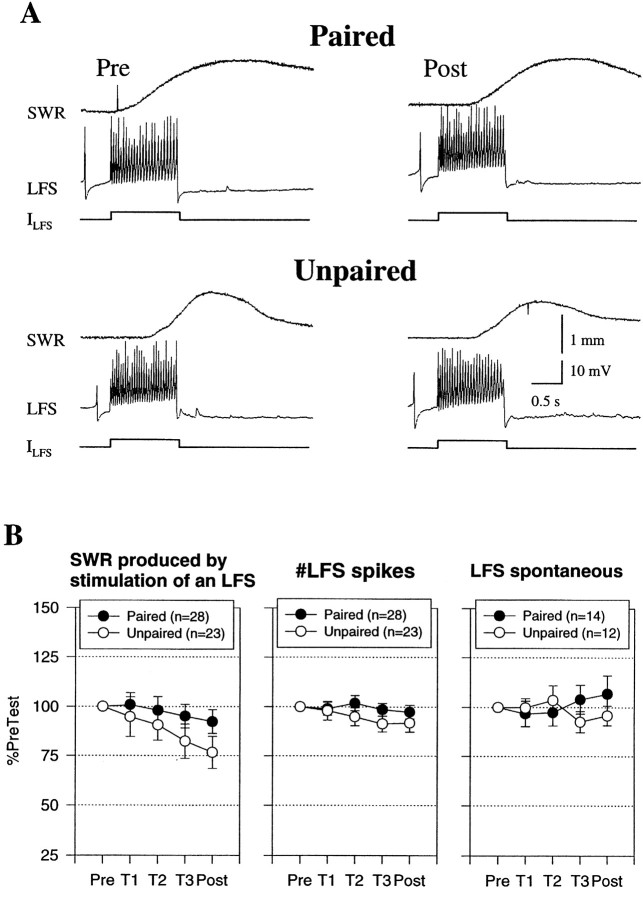
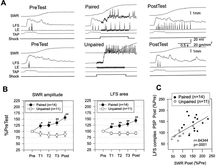
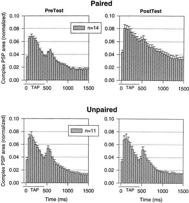
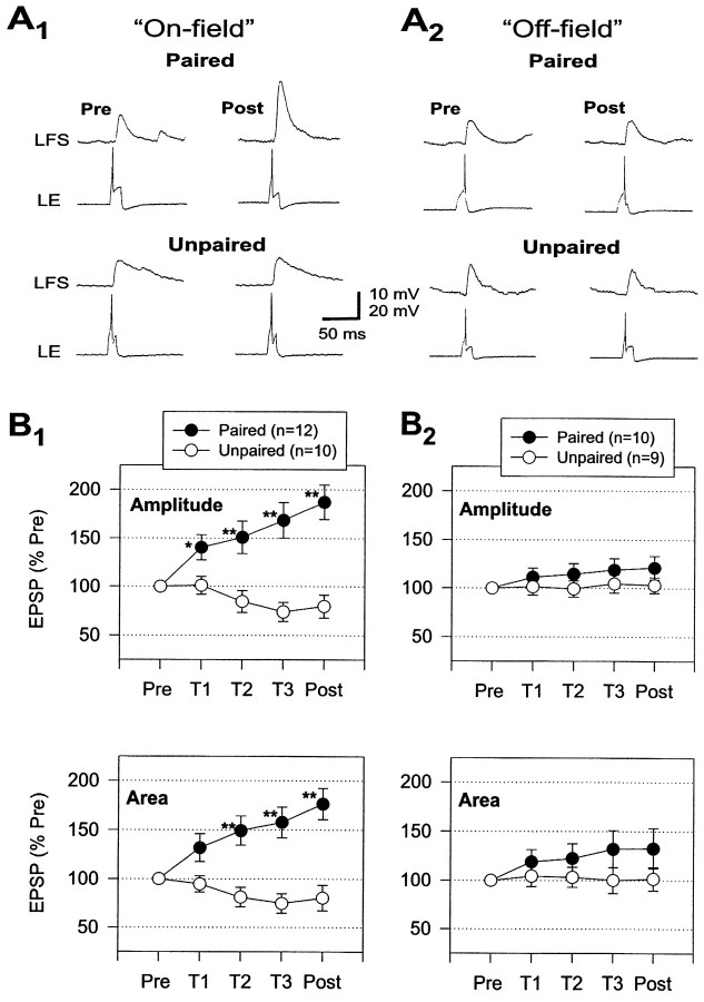
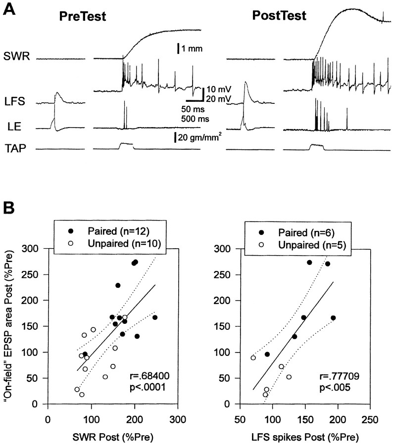
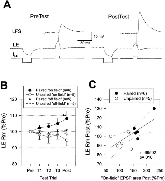
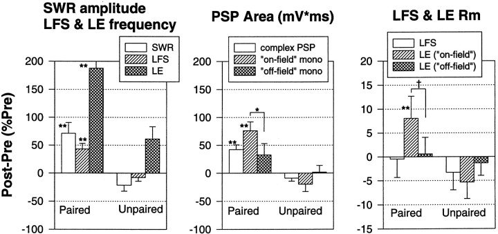
Plasticity at central synapses has long been thought to be the most likely mechanism for learning and memory, but testing that idea experimentally has proven to be difficult. For this reason, we have developed a simplified preparation of the Aplysia siphon withdrawal reflex that allows one to examine behavioral learning and memory while simultaneously monitoring synaptic connections between individual identified neurons in the CNS. We previously found that monosynaptic connections from LE siphon sensory neurons to LFS siphon motor neurons make a substantial contribution to the reflex in the siphon withdrawal preparation (Antonov et al., 1999a). We have now used that preparation to assess the contribution of various cellular mechanisms to classical conditioning of the reflex with a siphon tap conditioned stimulus (CS) and tail shock unconditioned stimulus (US). We find that, compared with unpaired training, paired training with the CS and US produces greater enhancement of siphon withdrawal and evoked firing of LFS neurons, greater facilitation of the complex PSP elicited in an LFS neuron by the siphon tap, and greater facilitation of the monosynaptic PSP elicited by stimulation of a single LE neuron. Moreover, the enhanced facilitation of monosynaptic LE-LFS PSPs is greater for LE neurons that fire during the siphon tap and correlates significantly with the enhancement of siphon withdrawal and evoked firing of the LFS neurons. These results provide the most direct evidence to date that activity-dependent plasticity at specific central synapses contributes to behavioral conditioning and support the idea that synaptic plasticity is a mechanism of learning and memory more generally.


Figures

Fig. 1.
Fig. 1.
Classical conditioning of theAplysia siphon withdrawal reflex (SWR) in the simplified preparation. A, Experimental preparation. G., Ganglion; S.N., sensory neuron; M.N., motor neuron. B, Protocol for paired training. See Materials and Methods for details.ITI, Intertrial interval. C, Examples of the siphon withdrawal produced by a tap to the siphon (the CS) on the pretest and final post-test after either paired (P) training, unpaired (UP) training, training with the CS alone, or training with the US (tail shock) alone. D, Average results in experiments such as those shown in C. Paired training produced a greater increase in the amplitude of siphon withdrawal than unpaired training, training with the CS alone, or training with the US alone. *p < 0.05; **p < 0.01 compared with paired. Responses have been normalized to the average values on the pretest, which were 3.2 mm (paired), 2.7 mm (unpaired), 2.7 mm (CS alone), and 3.1 mm (US alone), not significantly different by a one-way ANOVA. The average unconditioned responses to the first tail shock were 7.5 mm (paired), 5.9 mm (unpaired), and 7.8 mm (US alone), also not significantly different.
Fig. 2.
Fig. 2.
Increase in evoked firing of LFS siphon motor neurons and LE siphon sensory neurons during classical conditioning.A, Examples of the behavioral and cellular responses to the conditioned stimulus on the pretest and the final post-test after paired and unpaired training. In some cases the spike amplitudes have been attenuated because of the limited frequency response of the recording. B, Average siphon withdrawal and evoked firing of LFS and LE neurons recorded in the same experiments. Paired training produced a greater increase in the amplitude of siphon withdrawal, which was accompanied by a greater increase in evoked firing of the LFS and LE neurons in the first 1 sec. *p < 0.05; **p < 0.01 compared with unpaired in this and subsequent figures. The average values on the pretest were 2.4 mm (paired) and 2.7 mm (unpaired) for siphon withdrawal, 13.7 Hz (paired) and 16.3 Hz (unpaired) for LFS firing, and 5.2 Hz (paired) and 3.9 Hz (unpaired) for LE firing, not significantly different by t tests. The average unconditioned responses to the first tail shock were 4.3 mm (paired) and 4.9 mm (unpaired), not significantly different. C, There was a significant correlation between the increase in siphon withdrawal and the increase in evoked firing of the LFS neurons. Thesolid line indicates the linear regression, and thedashed lines indicate the 95% confidence intervals for the regression. SWR, Siphon withdrawal reflex.
Fig. 3.
Fig. 3.
Average pattern of firing of LFS motor neurons and LE sensory neurons on the pretest and final post-test after paired and unpaired training in the same experiments as in Figure 2. The number of spikes in each 100 msec interval has been normalized to the total number of spikes on the pretest in each experiment. The average values on the pretest were 15.8 spikes (paired) and 18.8 spikes (unpaired) for LFS firing and 5.2 spikes (paired) and 3.9 spikes (unpaired) for LE firing, not significantly different. Thehorizontal bar below the x-axis indicates the approximate duration of the siphon tap.
Fig. 4.
Fig. 4.
Classical conditioning did not involve significant changes at sites distal to the synapses onto the motor neurons.A, Examples of siphon withdrawal produced by constant current intracellular stimulation of an LFS neuron (ILFS) 30 sec after the pretest and the final post-test after paired and unpaired training. B, Average siphon withdrawal and number of spikes produced by intracellular stimulation of an LFS neuron in experiments like those shown inA, as well as the spontaneous firing rate of the LFS neuron 5 sec before each test. There was no significant difference in siphon withdrawal and also no differences in excitability or spontaneous firing of the motor neuron. The average values on the pretest were 0.7 mm (paired) and 0.8 mm (unpaired) for siphon withdrawal, 23.1 spikes (paired) and 26.2 spikes (unpaired) for LFS spikes, and 1.6 Hz (paired) and 1.6 Hz (unpaired) for LFS spontaneous firing, not significantly different by ttests. SWR, Siphon withdrawal reflex.
Fig. 5.
Fig. 5.
Facilitation of the complex PSP in LFS motor neurons during classical conditioning. A, Examples of the complex PSP produced in an LFS motor neuron by the siphon tap on the pretest and the final post-test after paired and unpaired training. In these experiments, the motor neuron was hyperpolarized to approximately −90 mV for a few seconds to prevent it from firing during the tap. B, Average magnitudes of siphon withdrawal and the complex PSP recorded in the same experiments. Paired training produced a greater increase in the amplitude of siphon withdrawal that was accompanied by a greater increase in the area of the complex PSP in the first 1 sec after its onset. The average values on the pretest were 1.8 mm (paired) and 1.7 mm (unpaired) for siphon withdrawal, and 36,369 mVmsec (paired) and 34,516 mVmsec (unpaired) for PSP area, not significantly different by t tests. The average unconditioned responses to the first tail shock were 3.7 mm (paired) and 4.1 mm (unpaired), not significantly different.C, There was a significant correlation between the increase in the amplitude of siphon withdrawal and the increase in the area of the complex PSP.
Fig. 6.
Fig. 6.
The average shape of the complex PSP in LFS motor neurons on the pretest and the final post-test after paired and unpaired training in the same experiments as in Figure 5. The PSP in each 50 msec interval has been normalized to the total area on the pretest in each experiment. The average values on the pretest were 44,018 mVmsec (paired) and 41,132 mVmsec (unpaired), not significantly different.
Fig. 7.
Fig. 7.
Facilitation of the monosynaptic PSP from an LE neuron to an LFS neuron during classical conditioning.A, Examples of the monosynaptic PSP produced in an LFS neuron by intracellular stimulation of an on-field (A1) and off-field (A2) LE neuron ∼10 sec before the siphon tap on the pretest and the final post-test after paired and unpaired training. B, Average modulation of the monosynaptic PSPs during paired and unpaired training. Paired training produced a greater increase than unpaired training in the amplitude and area of PSPs from LE neurons that fired during the siphon tap (on-field; B1). The increase in PSPs from on-field LE neurons during paired training was also significantly greater than the increase in PSPs from LE neurons that did not fire during the siphon tap (off-field;B2). The average values on the pretest were 10.8 mV and 820 mVmsec (paired, on-field), 13.0 mV and 1040 mVmsec (unpaired, on-field), 10.6 mV and 633 mVmsec (paired, off-field), and 14.2 mV and 1031 mVmsec (unpaired, off-field), not significantly different by ANOVAs.
Fig. 8.
Fig. 8.
Correlations between on-field monosynaptic PSPs and siphon withdrawal or LFS firing during classical conditioning.A, Examples of the monosynaptic PSP recorded ∼10 sec before siphon withdrawal and evoked firing of an LFS neuron on the pretest and the final post-test in a single experiment.B, There were significant correlations between the increase in the area of on-field monosynaptic PSPs and the increases in the amplitude of siphon withdrawal or number of evoked spikes in an LFS motor neuron. SWR, Siphon withdrawal reflex.
Fig. 9.
Fig. 9.
Increase in input resistance of LE sensory neurons during classical conditioning. A, Examples of the input resistance of an LE neuron measured with a hyperpolarizing intracellular current pulse (ILE) ∼10 sec before recording the monosynaptic PSP on the pretest and the final post-test in a single experiment. B, Average change in input resistance in experiments like the one shown in A. Paired training produced a greater increase than unpaired training in the input resistance of on-field LE neurons. There were no significant changes in the input resistance of off-field LE neurons.C, There was a significant correlation between the increase in the area of the monosynaptic PSP and the increase in the input resistance of on-field LE neurons. Rm, Membrane resistance.
Fig. 10.
Fig. 10.
Summary of results of the electrophysiological experiments. The siphon withdrawal reflex (SWR) in the simplified preparation undergoes classical conditioning that is accompanied by pairing-specific increases in evoked firing of both LFS motor neurons and LE sensory neurons, the complex PSP in LFS neurons, the monosynaptic PSP from on-field LE neurons to LFS neurons, and the input resistance of on-field LE neurons (**p < 0.01; *p < 0.05; †p < 0.05, one-tailed compared with either unpaired or off-field). Moreover, there were significant correlations between most of these measures. These results provide direct evidence that conditioning is attributable in part to activity-dependent plasticity of monosynaptic sensory neuron–motor neuron PSPs.

References

    1. Antonov I, Kandel ER, Hawkins RD. The contribution of facilitation of monosynaptic PSPs to dishabituation and sensitization of the Aplysia siphon withdrawal reflex. J Neurosci. 1999a;19:10438–10450. - PMC - PubMed
    1. Antonov I, Antonova I, Hawkins RD. Activity-dependent facilitation of monosynaptic sensory neuron-motor neuron PSPs contributes to classical conditioning of the Aplysia siphon-withdrawal reflex in a simplified preparation. Soc Neurosci Abstr. 1999b;25:1129.
    1. Antonov I, Kandel ER, Hawkins RD. Contribution of pre- and postsynaptic mechanisms to activity-dependent facilitation during classical conditioning of the Aplysia siphon-withdrawal reflex. Soc Neurosci Abstr. 2000;26:1523.
    1. Bailey CH, Castellucci VF, Koester J, Kandel ER. Cellular studies of peripheral neurons in siphon skin of Aplysia californica. J Neurophysiol. 1979;42:530–557. - PubMed
    1. Bao J-X, Kandel ER, Hawkins RD. Involvement of pre- and postsynaptic mechanisms in posttetanic potentiation at Aplysia synapses. Science. 1997;275:969–973. - PubMed

Publication types