. 2001 Aug;13(8):1803-18.
doi: 10.1105/tpc.010116.

The PET1-CMS mitochondrial mutation in sunflower is associated with premature programmed cell death and cytochrome c release

Affiliations

The PET1-CMS mitochondrial mutation in sunflower is associated with premature programmed cell death and cytochrome c release

J Balk et al. Plant Cell. 2001 Aug.

Abstract

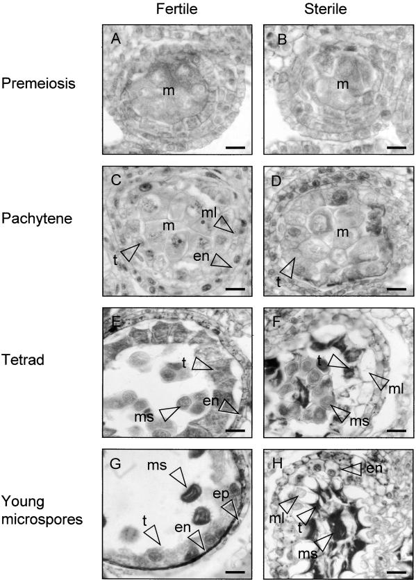
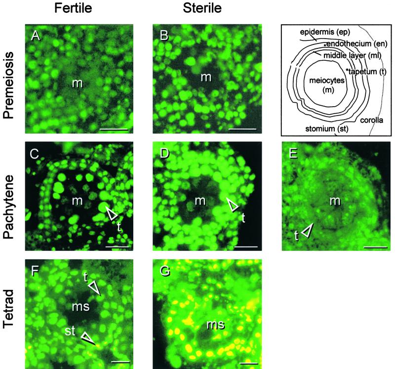
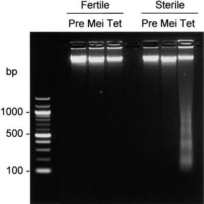
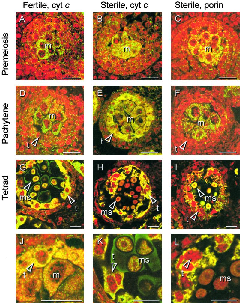
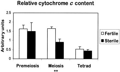
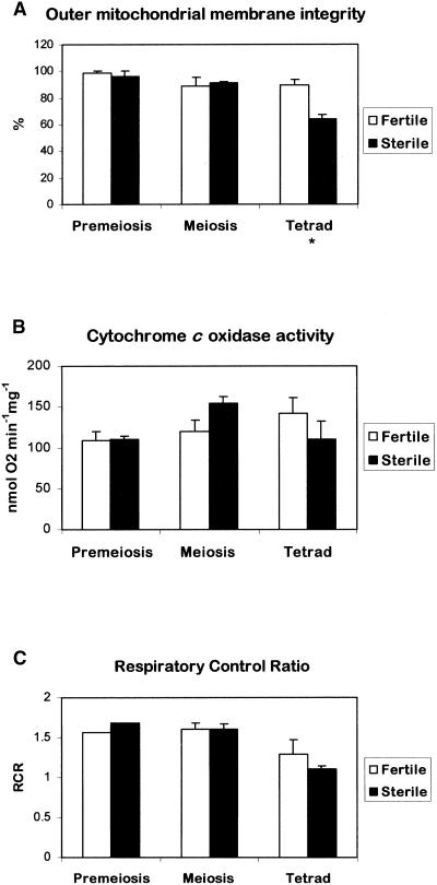
In mammals, mitochondria have been shown to play a key intermediary role in apoptosis, a morphologically distinct form of programmed cell death (PCD), for example, through the release of cytochrome c, which activates a proteolytic enzyme cascade, resulting in specific nuclear DNA degradation and cell death. In plants, PCD is a feature of normal development, including the penultimate stage of anther development, leading to dehiscence and pollen release. However, there is little evidence that plant mitochondria are involved in PCD. In a wide range of plant species, anther and/or pollen development is disrupted in a class of mutants termed CMS (for cytoplasmic male sterility), which is associated with mutations in the mitochondrial genome. On the basis of the manifestation of a number of morphological and biochemical markers of apoptosis, we have shown that the PET1-CMS cytoplasm in sunflower causes premature PCD of the tapetal cells, which then extends to other anther tissues. These features included cell condensation, oligonucleosomal cleavage of nuclear DNA, separation of chromatin into delineated masses, and initial persistence of mitochondria. In addition, immunocytochemical analysis revealed that cytochrome c was released partially from the mitochondria into the cytosol of tapetal cells before the gross morphological changes associated with PCD. The decrease in cytochrome c content in mitochondria isolated from male sterile florets preceded a decrease in the integrity of the outer mitochondrial membrane and respiratory control ratio. Our data suggest that plant mitochondria, like mammalian mitochondria, play a key role in the induction of PCD. The tissue-specific nature of the CMS phenotype is discussed with regard to cellular respiratory demand and PCD during normal anther development.


Figures

Figure 1.
Figure 1.
Anther Development in Florets of the Fertile and Sterile Sunflower Lines. Four stages of anther development in the fertile line and the corresponding stages of development in the sterile line were compared using histological staining. The images are of cross-sections through single locules. (A) Fertile line, premeiosis. (B) Sterile line, premeiosis. (C) Fertile line, pachytene. (D) Sterile line, pachytene. (E) Fertile line, tetrad stage. (F) Sterile line, tetrad stage. (G) Fertile line, stage 6 according to Horner (1977): young microspores. (H) Sterile line, stage 6 as in (G). en, endothecium cell(s); ep, epidermal cell(s); m, meiocytes; ml, middle layer; ms, microspore(s); t, tapetal cell(s). Bars = 20 μm.
Figure 2.
Figure 2.
DNA Fragmentation in Florets of the Fertile and Sterile Sunflower Lines. Three stages of anther development in the fertile line and the corresponding stages of development in the sterile line were compared with respect to nuclear DNA fragmentation using the TUNEL assay (bright green to yellow is positive stain). (A) Fertile line, premeiosis. (B) Sterile line, premeiosis. (C) Fertile line, pachytene. (D) Sterile line, pachytene. (E) As a negative control, the terminal deoxynucleotidyl transferase enzyme was omitted from the labeling mixture. Sterile line, pachytene. (F) Fertile line, tetrad stage. (G) Sterile line, tetrad stage. m, meiocyte(s); ms, microspores; st, stomium cells; t, tapetal cell(s). Bars = 20 μm.
Figure 3.
Figure 3.
Oligonucleosomal Cleavage of Total DNA in Florets of the Fertile and Sterile Sunflower Lines. Agarose gel electrophoresis of total DNA stained with ethidium bromide. The developmental stages correspond to those stages used for the TUNEL assay (Figure 2). Pre, premeiosis; Mei, meiosis; Tet, tetrad stage.
Figure 4.
Figure 4.
Characterization of the Monoclonal Antibodies Used for in Situ Labeling. Protein gel blot analysis using monoclonal antibodies against rat cytochrome c (7H8.2C12) and maize porin (PM035) on 30 μg of mitochondrial (Mit), chloroplast (Chl), and cytosolic (Cts) proteins from florets (mitochondria and cytosol) or leaves (chloroplast) of the fertile sunflower line.
Figure 5.
Figure 5.
Immunolocalization of Cytochrome c in Florets of the Fertile and Sterile Sunflower Lines. Three stages of anther development in the fertile line were compared with the corresponding stages in the sterile line with respect to the in situ localization of cytochrome c. Thin sections from PEG-embedded plant material were labeled with monoclonal antibodies against rat cytochrome c or, as depicted for the sterile line only, with monoclonal antibodies against porin, an integral outer mitochondrial membrane protein. The sections were secondarily labeled with a FITC-conjugated antibody (yellow/green), and nucleic acids were stained with propidium iodide (red) to facilitate identification of the developmental stage. (A), (B), and (C) Premeiosis. (D), (E), and (F) Pachytene. (G), (H), and (I) Tetrad stage. (A), (D), and (G) show localization of cytochrome c during anther development in the fertile line. (B), (E), and (H) show localization of cytochrome c during anther development in the sterile line. (C), (F), and (I) show localization of porin during anther development in the sterile line. (J) Subcellular localization of cytochrome c in the sterile line at pachytene shown at higher magnification. (K) Cytochrome c localization and propidium iodide staining in the fertile line at the tetrad stage shown at higher magnification. The arrowhead points to the chromatin of a tapetal cell. (L) Cytochrome c localization and propidium iodide staining in the sterile line at the tetrad stage shown at higher magnification. The arrowhead points to the chromatin of a dying tapetal cell. cyt c, cytochrome c; m, meiocyte(s); t, tapetal cell(s); ms, microspore(s). Bars = 20 μm.
Figure 6.
Figure 6.
Relative Cytochrome c Content in Isolated Mitochondria from Florets of the Fertile and Sterile Sunflower Lines. The relative amount of cytochrome c was determined by protein blot analysis and densitometry, working within a linear range of 20 to 40 μg of mitochondrial proteins as determined experimentally. The data are the average values of four independent experiments (±se), for each of which mitochondrial preparations were derived from four to eight sunflower heads. Statistical analysis was performed using Student's t test on sample means (** 0.001 < P ≤ 0.01).
Figure 7.
Figure 7.
Respiratory Properties of Isolated Mitochondria from Florets of the Fertile and Sterile Sunflower Lines. Standard oxygen electrode assays were used to determine outer membrane integrity (A), cytochrome c oxidase activity (B), and respiratory control ratio (C). The data are averages of two independent experiments (±se) of those used for Figure 6. Statistical analysis was performed using Student's t test on sample means (* 0.01 < P ≤ 0.05).
Figure 8.
Figure 8.
Quantification of Mitochondrial CPN60 in Several Sunflower Tissues. Thick sections of several sunflower tissues were labeled with an antibody against CPN60, and the fluorescent signal of the secondary antibody was quantified using confocal microscopy and optical sectioning. The relative CPN60 signal is expressed per cell (A) and per unit volume (B). The data are average values of at least three cells (±se).

References

    1. Balk, J., Leaver, C.J., and McCabe, P.F. (1999). Translocation of cytochrome c from the mitochondria to the cytosol occurs during heat-induced programmed cell death in cucumber plants. FEBS Lett. 463, 151–154. - PubMed
    1. Beers, E. (1997). Programmed cell death during plant growth and development. Cell Death Differ. 4, 649–661. - PubMed
    1. Bino, R., Suurs, L.C.J.M., De Hoop, S.J., Van der Neut, A., Van Went, J.L., and Van Marrewijk, G.A.M. (1986). Characterization of cytoplasmic male sterility in Petunia hybrida and Zea mays: Localization and activity of cytochrome c oxidase. Euphytica 35, 905–918.
    1. Bossy-Wetzel, E., Newmeyer, D.D., and Green, D.R. (1998). Mitochondrial cytochrome c release in apoptosis occurs upstream of DEVD-specific caspase activation and independently of mitochondrial transmembrane depolarization. EMBO J. 17, 37–49. - PMC - PubMed
    1. Cai, J., Yang, J., and Jones, D.P. (1998). Mitochondrial control of apoptosis: The role of cytochrome c. Biochim. Biophys. Acta 1366, 139–149. - PubMed

Publication types