. 2001 Aug;13(8):1929-43.
doi: 10.1105/tpc.010109.

Dynamic recruitment of Cdc2 to specific microtubule structures during mitosis

Affiliations

Dynamic recruitment of Cdc2 to specific microtubule structures during mitosis

M Weingartner et al. Plant Cell. 2001 Aug.

Abstract

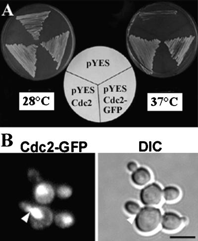
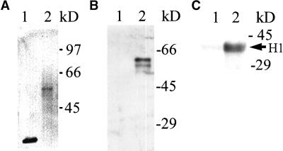
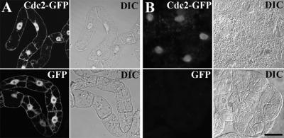
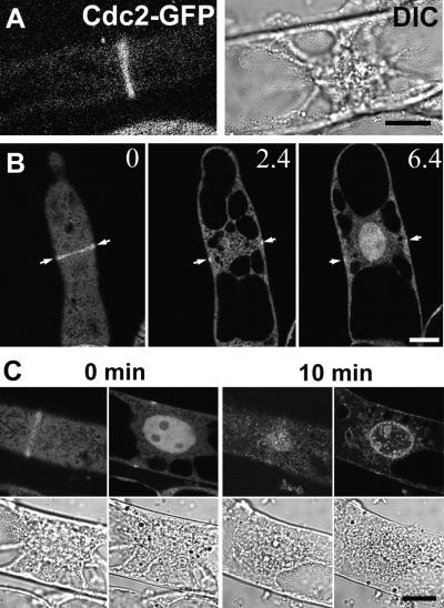
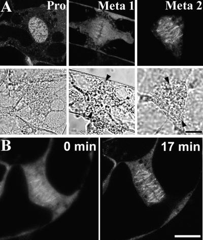
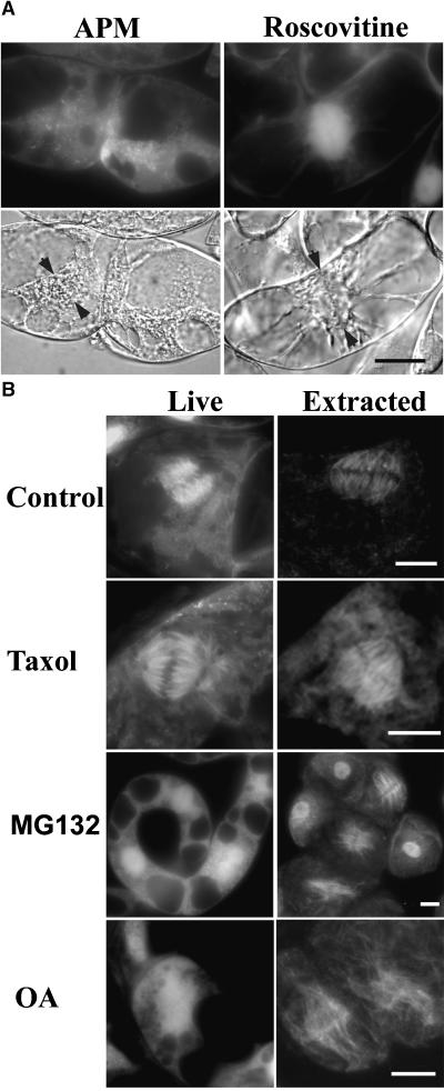
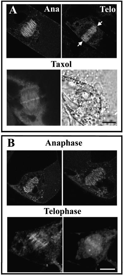
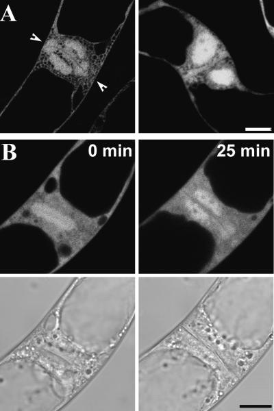
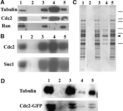
A-type cyclin-dependent kinases (CDKs), also known as cdc2, are central to the orderly progression of the cell cycle. We made a functional Green Fluorescent Protein (GFP) fusion with CDK-A (Cdc2-GFP) and followed its subcellular localization during the cell cycle in tobacco cells. During interphase, the Cdc2-GFP fusion protein was found in both the cytoplasm and the nucleus, where it was highly resistant to extraction. In premitotic cells, a bright and narrow equatorial band appeared on the cell surface, resembling the late preprophase band, which disintegrated within 10 min as followed by time-lapse images. Cdc2-GFP was not found on prophase spindles but left the chromatin soon after this stage and associated progressively with the metaphase spindle in a microtubule-dependent manner. Arresting cells in mitosis through the stabilization of microtubules by taxol further enhanced the spindle-localized pool of Cdc2-GFP. Toward the end of mitosis, Cdc2-GFP was found at the midzone of the anaphase spindle and phragmoplast; eventually, it became focused at the midline of these microtubule structures. In detergent-extracted cells, the Cdc2-GFP remained associated with mitotic structures. Retention on spindles was prevented by pretreatment with the CDK-specific inhibitor roscovitine and was enhanced by the protein phosphatase inhibitor okadaic acid. Furthermore, we demonstrate that both the endogenous CDK-A and Cdc2-GFP were cosedimented with taxol-stabilized plant microtubules from cell extracts and that Cdc2 activity was detected together with a fraction of polymerized tubulin. These data provide evidence that the A-type CDKs associate physically with mitotic structures in a microtubule-dependent manner and may be involved in regulating the behavior of specific microtubule arrays throughout mitosis.


Figures

Figure 1.
Figure 1.
Complementation Assay in Yeast. (A) Complementation analysis of the Saccharomyces cerevisiae CDC28-1N temperature-sensitive mutation containing the empty pYES yeast shuttle vector, pYES with Medsa;CDK-A;2 (pYES Cdc2), or the fusion of Medsa;CDK-A;2 with GFP (pYES Cdc2-GFP) and grown at 28 and 37°C on induction medium containing galactose. (B) Localization of Cdc2-GFP expressed in yeast cells at different phases of the cell cycle as shown by a fluorescence image (left) and by a differential interference contrast (DIC) image to visualize the cells (right). The arrowhead indicates a bud neck, where a spindle is localized in yeast. Bar = 5 μm.
Figure 2.
Figure 2.
Cdc2-GFP Fusion Protein Is Expressed in a Correct Size, Binds to p13suc1, and Is Active in Tobacco Cells. (A) Immunoblotting using extracts prepared from suspension cultured cells expressing GFP (lane 1) or Cdc2-GFP (lane 2) with a GFP-specific antibody. (B) Binding of GFP (lane 1) or Cdc2-GFP (lane 2) to p13suc1. Extracts prepared as in (A) were mixed with p13suc1 beads, and the bound fractions were immunoblotted with a GFP-specific antibody. (C) Histone H1 kinase activity of GFP (lane 1) and Cdc2-GFP (lane 2) immunopurified from extracts using a GFP-specific antibody. The phosphorylated histone H1 is indicated by an arrow. Molecular mass markers are labeled at right.
Figure 3.
Figure 3.
Cdc2-GFP Is Specifically Localized and Retained in the Nucleus. (A) Fluorescent microscopic images of Cdc2-GFP and GFP (left) and the corresponding DIC images (right) in live tobacco cells. (B) Fluorescent microscopic images of Cdc2-GFP and GFP cells extracted with 0.1% Triton X-100 (left) and the corresponding DIC images (right). Bar = 20 μm.
Figure 4.
Figure 4.
Cdc2-GFP Is Localized to a PPB-Like Structure. (A) Fluorescent microscopic image of a cell with a band-like structure (left) and the corresponding DIC image showing characteristic features of a cell in late G2-phase, such as the centrally positioned nucleus with condensing chromatin (right). Bar = 10 μm. (B) Optical sections of a cell having a PPB-like structure circumnavigating the cell. Three focal planes of 0, 2.4, and 6.4 μm are shown from images taken with a confocal laser scanning microscope. Bar = 10 μm. (C) Time-lapse images showing Cdc2-GFP during preprophase progression for a 10-min interval. The corresponding DIC images are shown underneath. Bar = 10 μm.
Figure 5.
Figure 5.
Cdc2-GFP Relocates from Chromatin to the Metaphase Spindle. (A) Fluorescent microscopic images of Cdc2-GFP in a prophase cell (Pro) and two metaphase cells (Meta 1 and Meta 2). The corresponding DIC images are shown underneath. Bar = 10 μm. (B) Time-lapse images showing Cdc2-GFP during metaphase progression for a 17-min interval. Bar = 10 μm.
Figure 6.
Figure 6.
Pharmacological Study of Cdc2-GFP Association with the Mitotic Spindle. (A) Cdc2-GFP fluorescence in metaphase cells treated with 10 μM of the microtubule-depolymerizing drug amiprophos methyl (APM) or with 100 μM of the CDK inhibitor roscovitine. The corresponding DIC images are shown underneath. Arrowheads indicate the metaphase plates. Bar = 10 μm. (B) Cdc2-GFP fluorescence in control metaphase cells and cells treated with 50 μM taxol, 100 μM MG132, and 0.2 μM okadaic acid (OA). Living cells are shown at left and cells extracted with 0.1% Triton X-100 are shown at right. Bars = 10 μm.
Figure 7.
Figure 7.
Cdc2-GFP Localization during Anaphase and Telophase. (A) Cdc2-GFP fluorescence in anaphase (Ana) and telophase (Telo) cells is shown at top. Arrows indicate the accumulation of Cdc2-GFP signal on the midline of the phragmoplast in a telophase cell. The bottom images show an anaphase cell treated with taxol (left, fluorescent image; right, corresponding DIC image). The accumulation of Cdc2-GFP along the midline of the anaphase spindle was pronounced in taxol-treated cells. Bar = 10 μm. (B) Time-lapse images showing Cdc2-GFP relocalization during 30-min intervals in an anaphase cell (top) and a telophase cell (bottom). Bar = 10 μm.
Figure 8.
Figure 8.
Reassociation of Cdc2-GFP with Chromatin in Late Telophase. (A) Cdc2-GFP fluorescence is shown in two late telophase cells. Arrowheads indicate the forming cell wall. Bar = 10 μm. (B) Time-lapse fluorescent images of Cdc2-GFP in a late telophase cell during a 25-min interval. The corresponding DIC images are shown underneath. Bar = 10 μm.
Figure 9.
Figure 9.
CDK and Cdc2-GFP Are Associated with Polymerized Microtubules in Microtubule Spin-Down Experiments. (A) Protein gel blot analysis of protein extracts from V. faba root tips probed with anti-tubulin, anti-Cdc2, and anti-Ran antibodies. Lane 1, total cell extract; lanes 2 and 3, the pellet and supernatant, respectively, of total extract after high speed centrifugation in the absence of taxol; lanes 4 and 5, as in lanes 2 and 3 but with taxol. (B) Histone H1 kinase activities in the same samples as in (A). CDK was purified from these samples by immunoprecipitation with an anti-Cdc2 antibody or by binding to p13suc1 and loaded in the same order as in (A). Histone H1 was used as a substrate. (C) Silver-stained SDS-polyacrylamide gel of samples as described in (A). Molecular mass markers from top to bottom are 212, 116, 97.4, 66.2, 57.5, and 40 kD. The position of tubulin is marked with an arrowhead. (D) Cdc2-GFP from tobacco cells is precipitated in a taxol-dependent manner. Samples were prepared and loaded in a similar manner as in (A) from suspension-cultured tobacco cells expressing Cdc2-GFP. Results of immunoblot analysis with anti-tubulin and anti–Cdc2-GFP antibodies are shown.

Similar articles

Cited by

References

    1. Alfa, C.E., Ducommun, B., Beach, D., and Hyams, J.S. (1990). Distinct nuclear and spindle pole body population of cyclin-cdc2 in fission yeast. Nature 347, 680–682. - PubMed
    1. Asada, T., Kuriyama, R., and Shibaoka, H. (1997). TKRP125, a kinesin-related protein involved in the centrosome-independent organization of the cytokinetic apparatus in tobacco BY-2 cells. J. Cell Sci. 110, 179–189. - PubMed
    1. Ayaydin, F., Vissi, E., Meszaros, T., Miskolczi, P., Kovacs, I., Feher, A., Dombradi, V., Erdodi, F., Gergely, P., and Dudits, D. (2000). Inhibition of serine/threonine-specific protein phosphatases causes premature activation of cdc2MsF kinase at G2/M tran-sition and early mitotic microtubule organisation in alfalfa. Plant J. 23, 85–96. - PubMed
    1. Bailly, E., Doree, M., Nurse, P., and Bornens, M. (1989). p34cdc2 is located in both nucleus and cytoplasm; part is centrosomally associated at G2/M and enters vesicles at anaphase. EMBO J. 8, 3985–3995. - PMC - PubMed
    1. Barroso, C., Chan, J., Allan, V., Doonan, J., Hussey, P., and Lloyd, C. (2000). Two kinesin-related proteins associated with the cold-stable cytoskeleton of carrot cells: Characterization of a novel kinesin, DcKRP120–2. Plant J. 24, 859–868. - PubMed

Publication types

MeSH terms

LinkOut - more resources