. 2001 Aug;13(8):1945-57.
doi: 10.1105/tpc.010084.

Peroxisomal metabolic function is required for appressorium-mediated plant infection by Colletotrichum lagenarium

Affiliations

Peroxisomal metabolic function is required for appressorium-mediated plant infection by Colletotrichum lagenarium

A Kimura et al. Plant Cell. 2001 Aug.

Abstract

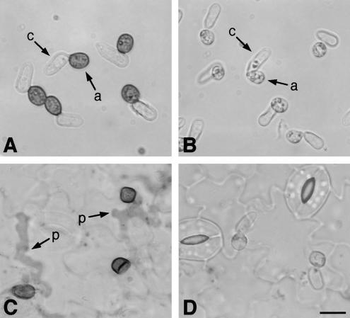
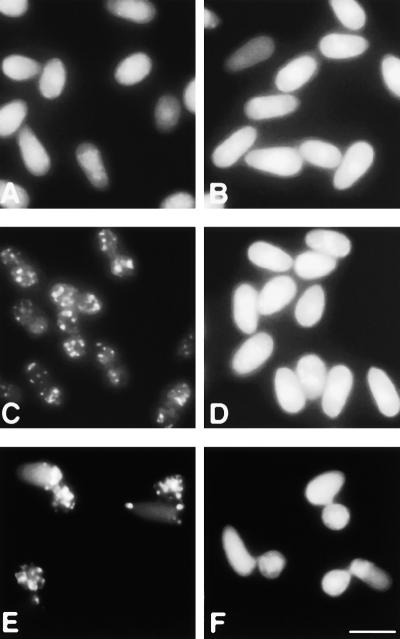
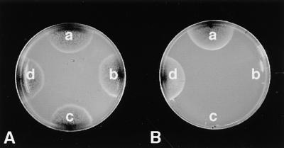
Peroxisomes are organelles that perform a wide range of metabolic functions in eukaryotic cells. However, their role in fungal pathogenesis is poorly understood. Here we report that ClaPEX6, an ortholog of PEX6, is required for the fungus Colletotrichum lagenarium to infect host plants. ClaPEX6 was identified in random insertional mutagenesis experiments aimed at elucidating genes involved in pathogenesis. Functional analysis, using a green fluorescent protein cassette containing the peroxisomal targeting signal1 (PTS1), revealed that import of PTS1-containing proteins is impaired in clapex6 mutants generated by targeted gene disruption. Failure of growth on fatty acids shows an inability of fatty acid beta-oxidation in these mutants. These results indicate that disruption of ClaPEX6 impairs peroxisomal metabolism, even though clapex6 mutants show normal growth and conidiation in nutrient-rich conditions. The clapex6 mutants formed small appressoria with severely reduced melanization that failed to form infectious hyphae. These data indicate that peroxisomes are necessary for appressorium-mediated penetration of host plants. The addition of glucose increased the pathogenicity of clapex6 mutants, suggesting that the glucose metabolic pathway can compensate partially for peroxisomes in phytopathogenicity.


Figures

Figure 1.
Figure 1.
DNA Gel Blot Analysis of the Nonpathogenic Mutant X86 and Strains Transformed with the Rescued Plasmid of X86. Genomic DNA from the wild-type strain 104-T (lane 1), X86 (lane 2), and four transformants of the wild type with the rescued plasmid (lanes 3 to 6) was digested with EcoRI, separated on a 1% agarose gel, and blotted. The blot was probed with a 1075-bp HindIII-ApaI genome fragment in the rescued plasmid. A single 7.0-kb EcoRI fragment was detected in the wild-type strain (arrow). In strain X86, the 7.0-kb fragment detected in the wild type disappeared but a 5.0-kb fragment was observed. Among the transformants with the rescued plasmid, two transformants (lanes 3 and 4) that lost pathogenicity did not contain the 7.0-kb fragment but contained the 5.0-kb fragment, which was common with X86. On the other hand, the other two transformants (lanes 5 and 6) that showed pathogenicity maintained the 7.0-kb fragment.
Figure 2.
Figure 2.
Predicted Amino Acid Sequence of the C. lagenarium ClaPEX6 Gene. The deduced amino acid sequence of C. lagenarium ClaPEX6 (C.l.) was aligned with Pex6 proteins from P. chrysogenum (P.c.), and S. cerevisiae (S.c.) by using the CLUSTAL W program (Thompson et al., 1994). Similar residues are shown on gray backgrounds. Gaps introduced for alignment are indicated by dashes. The ClaPex6 sequence contains two Walker A and Walker B motifs and one AAA-protein family signature motif. The plasmid insertion point in ClaPEX6 of strain X86 is indicated by the arrowhead. The sequence of ClaPEX6 has been deposited in GenBank (accession number AF343063).
Figure 3.
Figure 3.
Gene Disruption of ClaPEX6. (A) ClaPEX6 locus and gene replacement vector. By homologous recombination through double crossing over, the 2.8-kb region of ClaPEX6 was replaced by a hph cassette. A, ApaI; B, BamHI; P, PstI. (B) DNA gel blot analysis of ClaPEX6 gene replacement mutants. Genomic DNA was isolated from the wild-type strain 104-T (lane 1), gene replacement transformants DPE1, DPE8, and DPE9 (lanes 2 to 4), and ectopic transformants DPE4 and DPE7 (lanes 5 and 6). Genomic DNA was digested with BamHI. The blot was hybridized with a 1.8-kb PstI fragment of the ClaPEX6 locus, indicated by the gray bar in (A).
Figure 4.
Figure 4.
Pathogenicity Assay of clapex6Δ Mutants. Conidial suspensions of tested strains were spotted on the right half of detached cucumber leaves. On the left half of the leaves, the wild-type strain was inoculated as a positive control. (A) clapex6 deletion mutant DPE1. (B) Ectopic transformant DPE4.
Figure 5.
Figure 5.
Appressoria Formed by clapex6 Mutants and Their Function. (A) and (B) Conidial suspensions of the wild type and clapex6 mutant, respectively, were spotted on glass slides and incubated for 12 hr. Appressoria of the clapex6 mutant are smaller and show severe reduction of melanization. (C) and (D) Conidia were inoculated on cucumber cotyledons, incubated for 4 days, and stained with lactophenol aniline blue. (A) and (C) show the wild-type strain 104-T; (B) and (D) show the clapex6 mutant DPE1. a, appressorium; c, conidium; p, penetration hyphae. Bar in (D) = 10 μm for (A) to (D).
Figure 6.
Figure 6.
Ultrastructure of Conidia of C. lagenarium. Conidia of the wild-type strain of C. lagenarium were fixed with 5% KMnO4 and processed for electron microscopy. The arrowhead indicates the peroxisome. L, lipid body; M, mitochondrion; N, nucleus; V, vacuole. Bar = 1 μm.
Figure 7.
Figure 7.
Intracellular Localization of the GFP-PTS1 Fusion Protein in the clapex6 Mutant. (A) and (B) Green fluorescence of the intact GFP gene expressed in preincubated conidia of the wild type and the clapex6 mutant. (C) and (D) Subcellular localization of GFP-PTS1 expressed in preincubated conidia. (E) and (F) Subcellular localization of GFP-PTS1 in appressoria. (A), (C), and (E) show the wild-type strain; (B), (D), and (F) show the clapex6 mutant. Bar in (F) = 10 μm for (A) to (F).
Figure 8.
Figure 8.
Growth of clapex6 Mutants on Fatty Acids. Growth ability of the wild type (104-T), the clapex6 mutants (DPE1 and DPE8), and the ectopic transformant (DPE4) was investigated on glucose medium and fatty acid medium as a sole carbon source. Fatty acid medium contains 0.5% Tween 80. The clapex6 mutants failed to grow on the fatty acid medium. Plate A contained glucose medium; plate B contained fatty acid medium. a, wild-type strain; b, DPE1; c, DPE8; d, DPE4.
Figure 9.
Figure 9.
Partial Restoration of the Appressorium Phenotype in clapex6 Mutants by the Addition of Glucose and an Intermediate Product of Melanin. Conidia of the clapex6 mutant were incubated for 12 hr at 24°C in water, glucose solution, or melanin intermediate (scytalone) solution. (A) Water. (B) 1 mM glucose. (C) 1 mM scytalone. Bar in (C) = 10 μm for (A) to (C).

Similar articles

Cited by

References

    1. Adachi, K., and Hamer, J.E. (1998). Divergent cAMP signaling pathways regulate growth and pathogenesis in the rice blast fungus Magnaporthe grisea. Plant Cell 10, 1361–1373. - PMC - PubMed
    1. Agrios, G.N. (1988). Plant Pathology, 3rd ed. (San Diego, CA: Academic Press).
    1. Baerends, R.J.S., Faber, K.N., Kiel, J.A.K.W., van der Klei, J., Harder, W., and Veenhuis, M. (2000). Sorting and function of peroxisomal membrane proteins. FEMS Microbiol. Rev. 24, 291–301. - PubMed
    1. Bechinger, C., Giebel, K.-F., Schnell, M., Leiderer, P., Deising, H.B., and Bastmeyer, M. (1999). Optical measurements of invasive forces exerted by appressoria of a plant pathogenic fungus. Science 285, 1896–1899. - PubMed
    1. Beevers, H. (1979). Microbodies in higher plants. Annu. Rev. Plant Physiol. 30, 159–193.

Publication types

MeSH terms

Substances

Associated data

LinkOut - more resources