

Production and distribution of interleukin 15 and its receptors (IL-15Ralpha and IL-R2beta) in the implant interface tissues obtained during revision of failed total joint replacement
- PMID: 11488993
- PMCID: PMC2517707
- DOI: 10.1046/j.1365-2613.2001.iep0082-0201-x
Production and distribution of interleukin 15 and its receptors (IL-15Ralpha and IL-R2beta) in the implant interface tissues obtained during revision of failed total joint replacement
Abstract
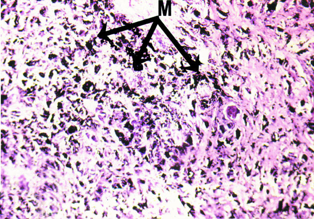
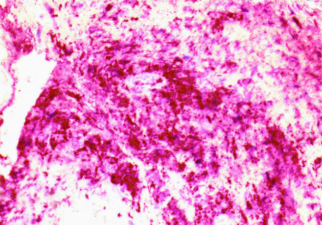
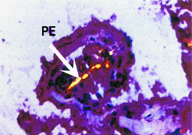
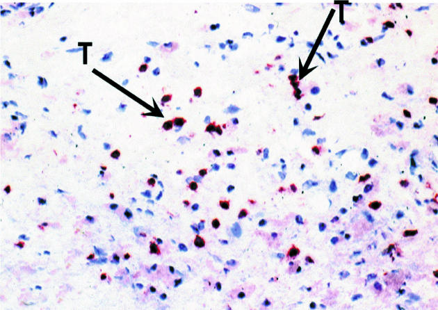
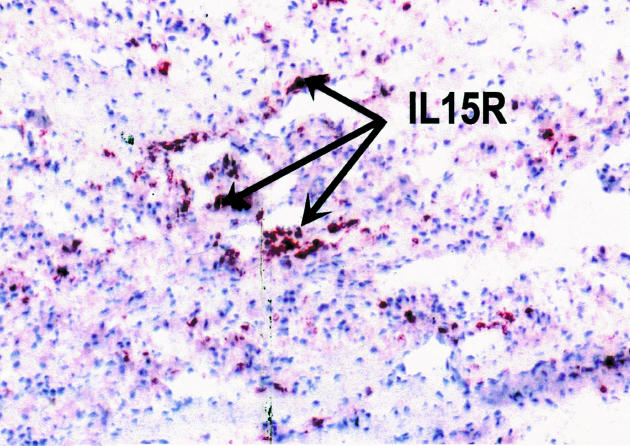
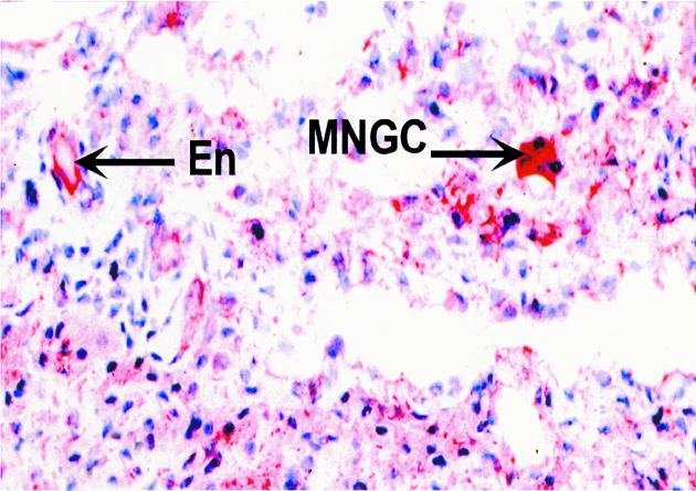
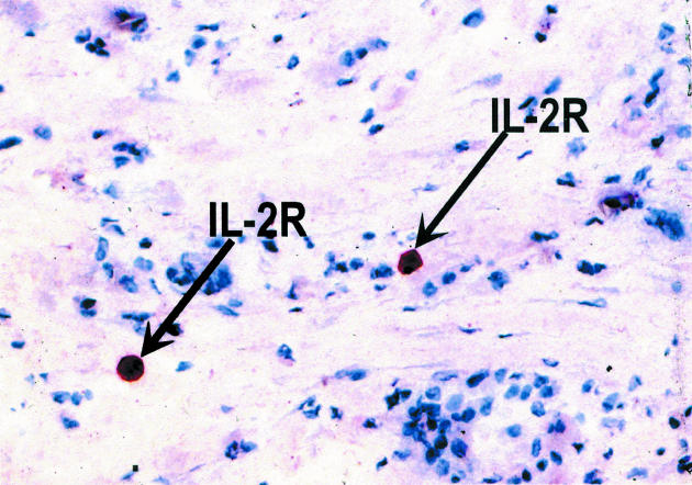
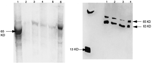
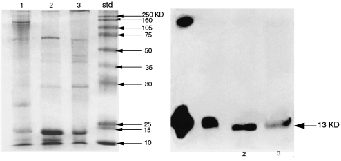
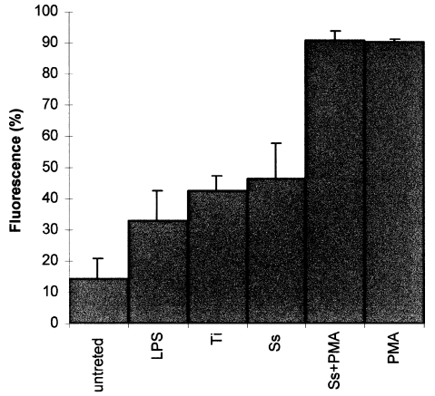
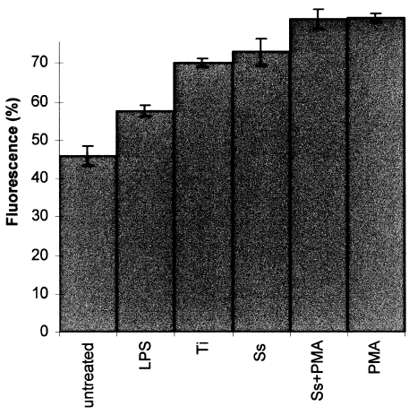
Failure of total joint replacement (TJR) is a major problem and it is estimated that 15-20% of TJR will fail within 5-10 years after implantation. Most TJR is attributed to aseptic loosening of the implants in association with resorption of related bone due to the release of bone-associated cytokines. IL-15 is a cytokine that activates T cells and natural killer (NK) cells. IL-15 protein is ubiquitous and is expressed in many tissues and cell types. Using immunohistochemical techniques, we demonstrated the expression of IL-15 and its receptors IL-15Ralpha and IL-2Rbeta in the interface tissues obtained from revision surgery. Both IL-15 protein and IL-15Ralpha were observed in macrophages, multinucleated giant cells and endothelial cells around blood vessels. Both the SDS-PAGE and western blot revealed multiple bands and after stages of glycosylation, this resulted in a band at 13 KDa which corresponds to the IL-15 protein. Again RT-PCR results demonstrated a band of 420 bp corresponding to the IL-15 protein. In addition, using U937 cells, the expression of both IL-15 protein and IL-15Ralpha were considerably up-regulated when challenged with retrieved metal particles. Our results illustrated the IL-15 to be an intact protein and that it is stored in the cytoplasm. A dye exclusion cell viability test displayed an increase in toxicity with an increase in the amount of metal particles added. There was a discrepancy between abundant IL-15 mRNA, intracellularly detectable IL-15 protein and apparently inefficient secretion. This suggests that IL-15 protein production is predominantly regulated post-transcriptionally and this is indicated by its strict regulation, especially at cell trafficking. Finally, unlike IL-2, IL-15 plays a certain role in bone resorption that leads to failed joint prostheses. It is apparent that this cytokine is an important T cell mediated immune response which needs further research.
Figures

References
-
- Bainbridge J, Dalby M, Curtis P, Knight M, Revell P. Cytoskeletal changes in macrophages and gaint cells following exposure to wear debris. 2000 Abstract submitted to the UK Society for Biometrials.
-
- Balknill F. Oxford: IRL Press; 1996. Cytokine: A practical approach.
-
- Bamford RN, Defilippis AP, Azimi N, Kurys G, Waldmann TA. The 5′ untranslated region, signal peptide and coding sequence of the carboxyl terminus of IL-15 participate in its multifaceted translational control. J. Immunol. 1998;160:4418–4426. - PubMed
-
- Daffada AAL, Murray EJ, Young SP. Control of activator protein 1 and nuclear factor Kappa β activity by international interleukin-6 and metals in HEPG2 cells. Biochim. Biophys. Acta. 1994;1222:234–240. - PubMed
-
- Doherty TM, Seder RA, Sher A. Induction and regulation of IL-15 expression in murine macrophages. J. Immunol. 1996;156:735–741. - PubMed
Publication types
MeSH terms
Substances
LinkOut - more resources
Full Text Sources
Other Literature Sources
Medical
