. 2001 Sep;183(17):5082-91.
doi: 10.1128/JB.183.17.5082-5091.2001.

LcrG-LcrV interaction is required for control of Yops secretion in Yersinia pestis

Affiliations

LcrG-LcrV interaction is required for control of Yops secretion in Yersinia pestis

J S Matson et al. J Bacteriol. 2001 Sep.

Abstract

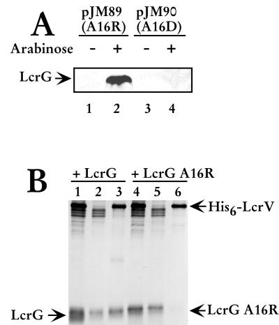
Yersinia pestis expresses a set of plasmid-encoded virulence proteins called Yops and LcrV that are secreted and translocated into eukaryotic cells by a type III secretion system. LcrV is a multifunctional protein with antihost and positive regulatory effects on Yops secretion that forms a stable complex with a negative regulatory protein, LcrG. LcrG has been proposed to block the secretion apparatus (Ysc) from the cytoplasmic face of the inner membrane under nonpermissive conditions for Yops secretion, when levels of LcrV in the cell are low. A model has been proposed to describe secretion control based on the relative levels of LcrG and LcrV in the bacterial cytoplasm. This model proposes that under secretion-permissive conditions, levels of LcrV are increased relative to levels of LcrG, so that the excess LcrV titrates LcrG away from the Ysc, allowing secretion of Yops to occur. To further test this model, a mutant LcrG protein that could no longer interact with LcrV was created. Expression of this LcrG variant blocked secretion of Yops and LcrV under secretion permissive conditions in vitro and in a tissue culture model. These results agree with the previously described secretion-blocking activity of LcrG and demonstrate that the interaction of LcrV with LcrG is necessary for controlling Yops secretion.


Figures

FIG. 1
FIG. 1
LcrG A16R is stably expressed in E. coli and does not interact with LcrV in Y. pestis. (A) pJM89 and pJM90 were transformed into E. coli strain Novablue cells. Overnight cultures were diluted 1:100 into fresh media and grown at 37°C for 1 h before the addition of 0.2% (wt/vol) arabinose to induce expression of LcrG A16R (lane 2) and LcrG A16D (lane 4). Samples were harvested by centrifugation after 3 h of growth with arabinose, and bacterial cells were permeabilized with Y-PER Reagent (Pierce) for 20 min. The cell debris was collected by centrifugation, and the supernatant was added to 2× sample buffer. Proteins were resolved by SDS-PAGE in a 15% polyacrylamide gel and analyzed by immunoblotting with α-LcrG. (B) Cells of Y. pestis KIM8-3002.8 (ΔlcrGV2) containing plasmids pAraG18K and pAra-HT-V (lanes 1 to 3) and Y. pestis KIM8-3002.8 (ΔlcrGV2) containing plasmids pJM89 and pAra-HT-V (lanes 4 to 6) were grown in TMH with calcium and induced with 0.2% (wt/vol) arabinose prior to the temperature shift to 37°C. Cultures were harvested after 4 h of growth at 37°C, and cellular extracts were prepared by disintegration in a French press (20,000 lb/in2). Low-speed centrifugation (14,000 × g) for 10 min removed unbroken cells and large debris. After centrifugation, the cleared extracts (lanes 1 and 4) were applied to a Talon column. Proteins that did not bind to the column were collected as the flowthrough fraction (lanes 2 and 5). Proteins eluted from the column with 50 mM imidazole were collected (lanes 3 and 6). Proteins were resolved by SDS-PAGE in a 13.5% polyacrylamide gel after dilution in 2X SDS sample buffer and analyzed by immunoblotting with α-LcrG and α-LcrV. Proteins were visualized by probing with alkaline phosphatase-conjugated secondary antibodies and developing with NBT-BCIP.
FIG. 2
FIG. 2
LcrG A16R blocks secretion regardless of the presence of calcium. (A) Cells of Y. pestis KIM8-3002 containing pBAD18-Kan (vector; lanes 1 and 2) and Y. pestis KIM8-3002.7 (ΔlcrG3) containing plasmids pBAD18-Kan (vector; lanes 3 and 4), pAraG (+LcrG; lanes 5 to 8), and pJM89 (+LcrG A16R; lanes 9 to 12) were grown in TMH with or without calcium. (B) Cells of Y. pestis KIM8-3002 containing pBAD18-Kan (vector; lanes 1 and 2) and Y. pestis KIM8-3002.6 (ΔlcrG2) containing plasmids pBAD18-Kan (vector; lanes 3 and 4), pAraG (+LcrG; lanes 5 to 8), and pJM89 (+LcrG A16R; lanes 9 to 12) were grown in TMH with or without calcium. Arabinose was added at 0.2% (wt/vol) to the cultures immediately prior to the temperature shift to 37°C to induce the expression of LcrG or LcrG A16R from the plasmids. Cultures were harvested after 4 h of growth at 37°C, and samples were fractionated into whole-cell and cell-free culture supernatants. Cell-free culture supernatant samples were separated by SDS-PAGE in a 12.5% polyacrylamide gel and analyzed by immunoblotting with an antibody cocktail containing α-LcrV, α-YopN, and α-YopE. Duplicate immunoblots were probed with α-YopM. Samples were separated by SDS-PAGE in a 15% polyacrylamide gel and analyzed by immunoblotting with α-LcrQ and α-LcrG on separate immunoblots. Proteins were visualized by probing with alkaline phospatase-conjugated secondary antibodies and developing with NBT-BCIP.
FIG. 3
FIG. 3
LcrG A16R does not fully repress expression of Yops. (A) Cells of Y. pestis KIM8-3002 containing pBAD18-Kan (vector; lanes 1 and 2) and Y. pestis KIM8-3002.7 (ΔlcrG3) containing plasmids pBAD18-Kan (vector; lanes 3 and 4), pAraG (+LcrG; lanes 5 to 8), and pJM89 (+LcrG A16R; lanes 9 to 12) were grown in TMH with or without calcium. (B) Cells of Y. pestis KIM8-3002 containing pBAD18-Kan (vector; lanes 1 and 2) and Y. pestis KIM8-3002.6 (ΔlcrG2) containing plasmids pBAD18-Kan (vector; lanes 3 and 4), pAraG (+LcrG; lanes 5 to 8), and pJM89 (+LcrG A16R; lanes 9 to 12) were grown in TMH with or without calcium. Arabinose was added at 0.2% (wt/vol) to the cultures prior to temperature shift to 37°C to induce the expression of LcrG or LcrG A16R from the plasmids. Cultures were harvested after 4 h of growth at 37°C, and samples were fractionated into whole-cell and cell-free culture supernatants. Whole-cell samples were separated by SDS-PAGE in a 12.5% polyacrylamide gel and analyzed by immunoblotting with an antibody cocktail containing α-LcrV, α-YopN, and α-YopE. Duplicate immunoblots were probed with α-YopM. Samples were separated by SDS-PAGE in a 15% polyacrylamide gel and analyzed by immunoblotting with α-LcrQ and α-LcrG on separate immunoblots. Proteins were visualized by probing with alkaline phospatase-conjugated secondary antibodies and developing with NBT-BCIP.
FIG. 4
FIG. 4
LcrG A16R blocks Y. pestis-induced cytotoxicity of HeLa cells. Y. pestis KIM8-3002 containing pBAD18-Kan, and KIM8-3002.7 (ΔlcrG3) containing pBAD18-Kan, pAraG, and pJM89 were used to infect HeLa cells at a multiplicity of infection of 5 to 10. Arabinose (ara) was added at 0.2% (wt/vol) to induce the expression of LcrG or LcrG A16R from the plasmids. After 4 h of expression, the cultures were viewed with Hoffman modulation optics to evaluate cytotoxicity and photographed with a green filter.
FIG. 5
FIG. 5
Phenotype of lcrGV. Cells of Y. pestis KIM8-3002 containing pBAD18-Kan (vector; lanes 1 and 2) and Y. pestis KIM8-3002.8 (ΔlcrGV2) containing plasmids pBAD18-Kan (vector; lanes 3 and 4), pAraG (+LcrG; lanes 5 and 6), pAraV (+LcrV; lanes 7 and 8), and pAraGV (+LcrGV; lanes 9 and 10) were grown in TMH with or without calcium. Arabinose was added at 0.2% (wt/vol) to each of the cultures immediately prior to the temperature shift to 37°C to induce the expression of LcrG, LcrV, or LcrG and LcrV from the plasmids. Cultures were harvested after 4 h of growth at 37°C, and samples were fractionated into whole-cell (A) and cell-free culture supernatants (B). Samples were separated by SDS-PAGE in a 12.5% polyacrylamide gel and analyzed by immunoblotting with an antibody cocktail containing α-LcrV, α-YopE, and α-LcrG. Duplicate immunoblots were probed with α-YopM. Proteins were visualized by probing with alkaline phospatase-conjugated secondary antibodies and developing with NBT-BCIP.
FIG. 6
FIG. 6
Model for LCR regulation in Y. pestis. In the presence of calcium, LcrG, YopN, and TyeA block the Ysc. LcrG is hypothesized to exert its blocking activity from the cytoplasm while YopN may block secretion at the cell surface. The combined secretion block retains LcrQ in the cell, resulting in repression of LCR-regulated genes (Repressed). In the absence of calcium or in the presence of eukaryotic cell contact, a block (possibly YopN) is released, allowing secretion of LcrQ ([Activation]). Secretion of LcrQ is believed to allow induction of LCR-regulated genes, including lcrV. Increased LcrV levels in the cytoplasm titrate LcrG away from the Ysc by forming a stable LcrG-LcrV complex. The removal of LcrG results in Yops and LcrV secretion and full induction of the LCR (Activated). If LcrG and LcrV are incapable of interacting, LcrG cannot be titrated away from the Ysc. This results in a constitutive blockage of Yops secretion regardless of calcium concentration or eukaryotic cell contact (LcrG-LcrV interaction blocked).

References

    1. Ausubel F M, Brent R, Kingston R E, Moore D D, Seidman J G, Smith J A, Struhl K, editors. Current protocols in molecular biology. New York, N.Y: John Wiley & Sons; 1998.
    1. Bergman T, Häkansson S, Forsberg Å, Norlander L, Macellaro A, Bäckman A, Bölin I, Wolf-Watz H. Analysis of the V antigen lcrGVH-yopBD operon of Yersinia pseudotuberculosis: evidence for a regulatory role of LcrH and LcrV. J Bacteriol. 1991;173:1607–1616. - PMC - PubMed
    1. Boland A, Sory M-P, Iriarte M, Kerbourch C, Wattiau P, Cornelis G R. Status of YopM and YopN in the Yersinia Yop virulon: YopM of Y. enterocolitica is internalized inside the cytosol of PU5–1.8 macrophages by the YopB, D, N delivery apparatus. EMBO J. 1996;15:5191–5201. - PMC - PubMed
    1. Boyd A P, Sory M-P, Iriarte M, Cornelis G R. Heparin interferes with translocation of Yop proteins into HeLa cells and binds to LcrG, a regulatory component of the Yersinia Yop apparatus. Mol Microbiol. 1998;27:425–436. - PubMed
    1. Cheng L W, Schneewind O. Yersinia enterocolitica TyeA, an intracellular regulator of the type III macinery, is required for specific targeting of YopE, YopH, YopM, and YopN into the cytosol of eukaryotic cells. J Bacteriol. 2000;182:3183–3190. - PMC - PubMed

LinkOut - more resources