. 2001 Aug 6;194(3):235-46.
doi: 10.1084/jem.194.3.235.

NTB-A [correction of GNTB-A], a novel SH2D1A-associated surface molecule contributing to the inability of natural killer cells to kill Epstein-Barr virus-infected B cells in X-linked lymphoproliferative disease

Affiliations

NTB-A [correction of GNTB-A], a novel SH2D1A-associated surface molecule contributing to the inability of natural killer cells to kill Epstein-Barr virus-infected B cells in X-linked lymphoproliferative disease

C Bottino et al. J Exp Med. .

Erratum in

  • J Exp Med 2001 Sep 3;194(5):following 703

Abstract

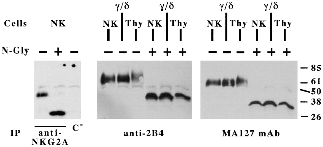
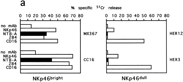
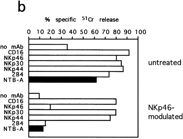
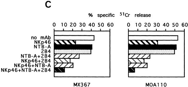
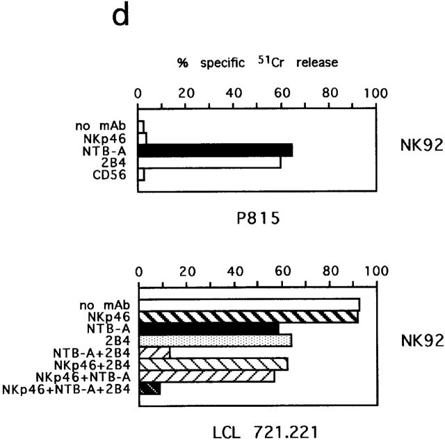
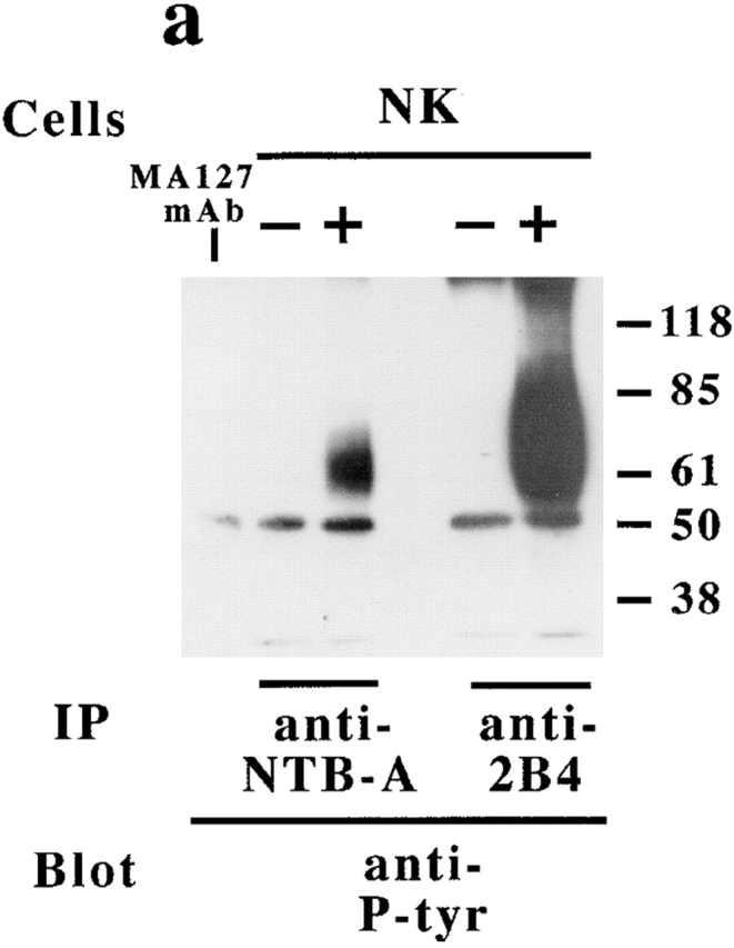
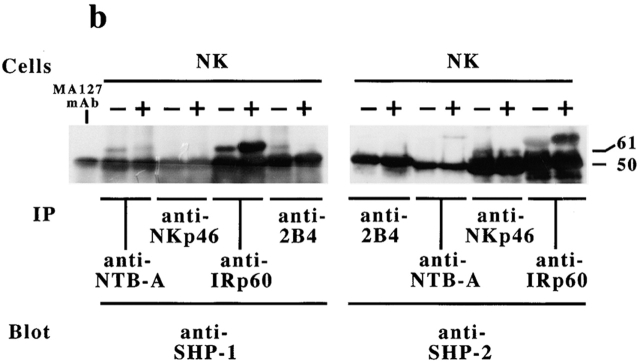
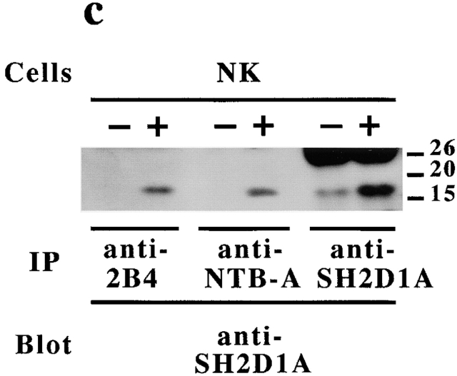
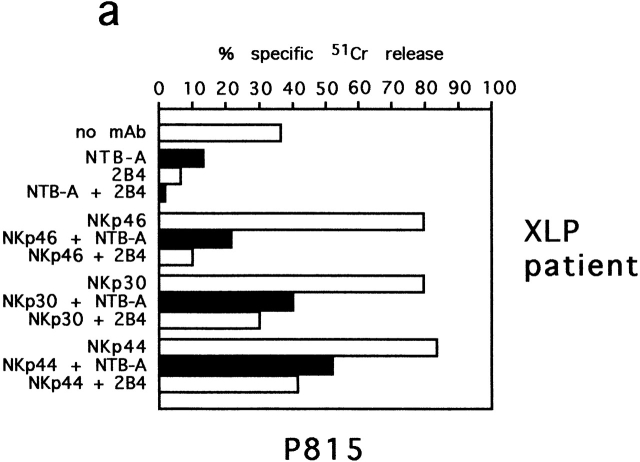
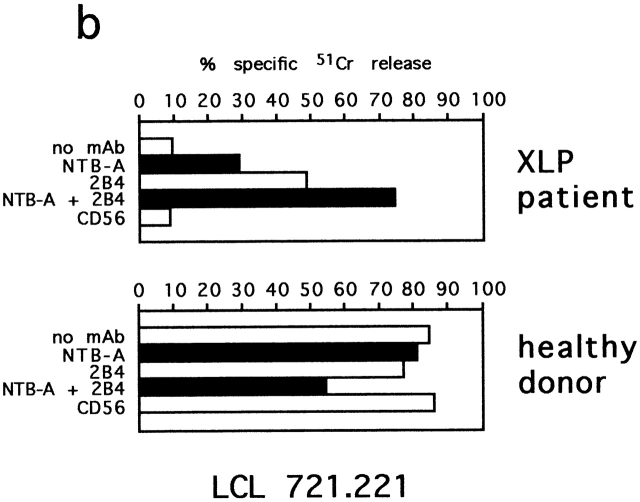
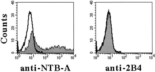
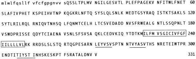
In humans, natural killer (NK) cell function is regulated by a series of receptors and coreceptors with either triggering or inhibitory activity. Here we describe a novel 60-kD glycoprotein, termed NTB-A, that is expressed by all human NK, T, and B lymphocytes. Monoclonal antibody (mAb)-mediated cross-linking of NTB-A results in the induction of NK-mediated cytotoxicity. Similar to 2B4 (CD244) functioning as a coreceptor in the NK cell activation, NTB-A also triggers cytolytic activity only in NK cells expressing high surface densities of natural cytotoxicity receptors. This suggests that also NTB-A may function as a coreceptor in the process of NK cell activation. Molecular cloning of the cDNA coding for NTB-A molecule revealed a novel member of the immunoglobulin superfamily belonging to the CD2 subfamily. NTB-A is characterized, in its extracellular portion, by a distal V-type and a proximal C2-type domain and by a cytoplasmic portion containing three tyrosine-based motifs. NTB-A undergoes tyrosine phosphorylation and associates with the Src homology 2 domain-containing protein (SH2D1A) as well as with SH2 domain-containing phosphatases (SHPs). Importantly, analysis of NK cells derived from patients with X-linked lymphoproliferative disease (XLP) showed that the lack of SH2D1A protein profoundly affects the function not only of 2B4 but also of NTB-A. Thus, in XLP-NK cells, NTB-A mediates inhibitory rather than activating signals. These inhibitory signals are induced by the interaction of NTB-A with still undefined ligands expressed on Epstein-Barr virus (EBV)-infected target cells. Moreover, mAb-mediated masking of NTB-A can partially revert this inhibitory effect while a maximal recovery of target cell lysis can be obtained when both 2B4 and NTB-A are simultaneously masked. Thus, the altered function of NTB-A appears to play an important role in the inability of XLP-NK cells to kill EBV-infected target cells.


Figures

Figure 1
Figure 1
Biochemical characterization of NTB-A molecules. NK cell, TCR-γ/δ1 cell and thymocyte (Thy) populations that had been surface labeled with biotin were immunoprecipitated (IP) with MA127 (anti–NTB-A) or S39 (anti-2B4) mAbs. Samples were treated (+) or not (−) with N glycosidase F and analyzed in a 9% SDS-PAGE under reducing conditions. Z270 (anti-NKG2A) and WT31 (C) (anti–TCR-α/β), both of IgG1 isotype, were used as controls. Molecular weight markers (kD) are indicated.
Figure 2
Figure 2
Functional analysis of NTB-A molecules in redirected killing assay and in NK-mediated killing of EBV+ target cells. (a) Representative NKp46bright (MX367, CC16) and NKp46dull (HER12, HER3) NK cell clones were analyzed in a redirected killing assay against (FcγR+) P815 murine target cells either in the absence or in the presence of mAbs specific for the indicated molecules. All the mAbs were of the IgG1 subclass. (b) The representative NKp46bright clone (MOA110) either untreated or modulated with anti-NKp46 (KL247, IgM) mAb was tested for cytolytic activity in a redirected killing assay against murine P815 target cells either in the absence or in the presence of mAbs specific for the indicated molecules. In both experiments the E/T ratio used was 8:1. (c) Two representative NKp46bright NK cell clones (MX367 and MOA110) were analyzed for cytolytic activity against the LCL 721.221 EBV cell line (HLA class ICD48+FcγR) either in the absence or in the presence of mAbs specific for the indicated molecules. The E/T ratio used was 2:1. (d) The NK92 NK cell line was analyzed for cytolytic activity against the (FcγR+) P815 murine target cells or the LCL 721.221 EBV cell line, either in the absence or in the presence of mAbs specific for the indicated molecules. The E/T ratio used was 5:1. The results are representative of seven independent experiments. The standard deviation of the mean of the triplicates was <4%.
Figure 2
Figure 2
Functional analysis of NTB-A molecules in redirected killing assay and in NK-mediated killing of EBV+ target cells. (a) Representative NKp46bright (MX367, CC16) and NKp46dull (HER12, HER3) NK cell clones were analyzed in a redirected killing assay against (FcγR+) P815 murine target cells either in the absence or in the presence of mAbs specific for the indicated molecules. All the mAbs were of the IgG1 subclass. (b) The representative NKp46bright clone (MOA110) either untreated or modulated with anti-NKp46 (KL247, IgM) mAb was tested for cytolytic activity in a redirected killing assay against murine P815 target cells either in the absence or in the presence of mAbs specific for the indicated molecules. In both experiments the E/T ratio used was 8:1. (c) Two representative NKp46bright NK cell clones (MX367 and MOA110) were analyzed for cytolytic activity against the LCL 721.221 EBV cell line (HLA class ICD48+FcγR) either in the absence or in the presence of mAbs specific for the indicated molecules. The E/T ratio used was 2:1. (d) The NK92 NK cell line was analyzed for cytolytic activity against the (FcγR+) P815 murine target cells or the LCL 721.221 EBV cell line, either in the absence or in the presence of mAbs specific for the indicated molecules. The E/T ratio used was 5:1. The results are representative of seven independent experiments. The standard deviation of the mean of the triplicates was <4%.
Figure 3
Figure 3
Analysis of the NTB-A–specific signal transduction pathway. (a) Cell lysates derived from a polyclonal NK cell population either untreated (−) or treated (+) with sodium pervanadate (100 μM, 10 min, 37°C), were sequentially immunoprecipitated (IP) with anti-2B4 and anti-NTBA mAbs. Samples were analyzed under reducing conditions in 8% SDS-PAGE and probed with anti-phosphotyrosine (anti-P-Tyr). (b) Cell lysates derived as in panel a were sequentially immunoprecipitated with anti-2B4, anti–NTB-A, anti-NKp46, and anti-IRp60. Equal amounts of each immunoprecipitate were analyzed under reducing conditions in 8% SDS-PAGE and probed with either anti–SHP-1 or anti–SHP-2 mAbs. (c) Cell lysates derived as in panel a were sequentially immunoprecipitated with anti-2B4, anti–NTB-A, and anti-SH2D1A. Samples were analyzed under reducing conditions in 14% SDS-PAGE and probed with anti-SH2D1A antiserum. In each panel molecular weight markers (kD) are indicated.
Figure 3
Figure 3
Analysis of the NTB-A–specific signal transduction pathway. (a) Cell lysates derived from a polyclonal NK cell population either untreated (−) or treated (+) with sodium pervanadate (100 μM, 10 min, 37°C), were sequentially immunoprecipitated (IP) with anti-2B4 and anti-NTBA mAbs. Samples were analyzed under reducing conditions in 8% SDS-PAGE and probed with anti-phosphotyrosine (anti-P-Tyr). (b) Cell lysates derived as in panel a were sequentially immunoprecipitated with anti-2B4, anti–NTB-A, anti-NKp46, and anti-IRp60. Equal amounts of each immunoprecipitate were analyzed under reducing conditions in 8% SDS-PAGE and probed with either anti–SHP-1 or anti–SHP-2 mAbs. (c) Cell lysates derived as in panel a were sequentially immunoprecipitated with anti-2B4, anti–NTB-A, and anti-SH2D1A. Samples were analyzed under reducing conditions in 14% SDS-PAGE and probed with anti-SH2D1A antiserum. In each panel molecular weight markers (kD) are indicated.
Figure 4
Figure 4
Functional analysis of NTB-A molecules in NK cells derived from XLP patients. (a) Polyclonal NK cell population derived from XLP patient A was analyzed for cytolytic activity in a redirected killing assay against the (FcγR+) P815 target cell line either in the absence or in the presence of mAbs specific for the indicated molecules. The E/T ratio used was 8:1. All the mAbs used in this experiment were of the IgG1 isotype. (b) Polyclonal NK cell populations derived from XLP patient A or from a healthy donor were analyzed for cytolytic activity against the LCL 721.221 EBV cell line (HLA class ICD48+FcγR) either in the absence or in the presence of mAbs specific for the indicated molecules. The E/T ratio used was 6:1. The results are representative of six independent experiments. The standard deviation of the mean of the triplicates was <5%.
Figure 4
Figure 4
Functional analysis of NTB-A molecules in NK cells derived from XLP patients. (a) Polyclonal NK cell population derived from XLP patient A was analyzed for cytolytic activity in a redirected killing assay against the (FcγR+) P815 target cell line either in the absence or in the presence of mAbs specific for the indicated molecules. The E/T ratio used was 8:1. All the mAbs used in this experiment were of the IgG1 isotype. (b) Polyclonal NK cell populations derived from XLP patient A or from a healthy donor were analyzed for cytolytic activity against the LCL 721.221 EBV cell line (HLA class ICD48+FcγR) either in the absence or in the presence of mAbs specific for the indicated molecules. The E/T ratio used was 6:1. The results are representative of six independent experiments. The standard deviation of the mean of the triplicates was <5%.
Figure 5
Figure 5
Surface expression of NTB-A in COS-7 cells transfected with the VR1012/KALI construct. COS-7 cells transfected with VR1012/KALI construct were stained with MA127 (anti–NTB-A) or PP35 (anti-2B4) mAbs followed by PE-conjugated goat anti–mouse second reagent and analyzed by flow cytometry. White profiles indicate cells incubated with the second reagent only.
Figure 6
Figure 6
Predicted amino acid sequence of NTB-A molecule. The putative signal peptide is indicated in lower case letters and the transmembrane region is boxed. Tyrosine-based motifs present in the cytoplasmic tail are underlined. The NTB-A nucleotide sequence is available from GenBank/EMBL/DDBJ under accession no. AJ277141.

References

    1. Moretta A., Bottino C., Vitale M., Pende D., Biassoni R., Mingari M.C., Moretta L. Receptors for HLA-class I molecules in human natural killer cells. Annu. Rev. Immunol. 1996;14:619–648. - PubMed
    1. Lanier L.L. NK cell receptors. Annu. Rev. Immunol. 1998;16:359–393. - PubMed
    1. Long E.O. Regulation of immune responses through inhibitory receptors. Annu. Rev. Immunol. 1999;17:875–904. - PubMed
    1. Moretta A., Bottino C., Vitale M., Pende D., Cantoni C., Mingari M.C., Biassoni R., Moretta L. Activating receptors and co-receptors involved in human natural killer cell-mediated cytolysis. Annu. Rev. Immunol. 2001;19:197–223. - PubMed
    1. Garrido F., Ruiz-Cabello F., Cabrera T., Pérez-Villar J.J., Lòpez-Botet M., Duggan-Keen M., Stern P.L. Implications for immunosurveillance of altered HLA-class I phenotypes in human tumours. Immunol. Today. 1997;18:89–95. - PubMed

Publication types

MeSH terms

Grants and funding