. 2001 Aug 6;194(3):285-300.
doi: 10.1084/jem.194.3.285.

Point mutation in essential genes with loss or mutation of the second allele: relevance to the retention of tumor-specific antigens

Affiliations

Point mutation in essential genes with loss or mutation of the second allele: relevance to the retention of tumor-specific antigens

G B Beck-Engeser et al. J Exp Med. .

Abstract

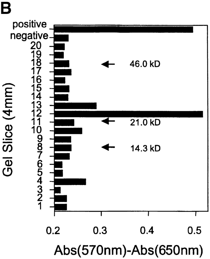
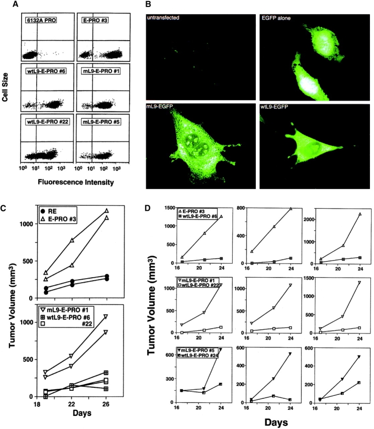
Antigens that are tumor specific yet retained by tumor cells despite tumor progression offer stable and specific targets for immunologic and possibly other therapeutic interventions. Therefore, we have studied two CD4(+) T cell-recognized tumor-specific antigens that were retained during evolution of two ultraviolet-light-induced murine cancers to more aggressive growth. The antigens are ribosomal proteins altered by somatic tumor-specific point mutations, and the progressor (PRO) variants lack the corresponding normal alleles. In the first tumor, 6132A-PRO, the antigen is encoded by a point-mutated L9 ribosomal protein gene. The tumor lacks the normal L9 allele because of an interstitial deletion from chromosome 5. In the second tumor, 6139B-PRO, both alleles of the L26 gene have point mutations, and each encodes a different tumor-specific CD4(+) T cell-recognized antigen. Thus, for both L9 and L26 genes, we observe "two hit" kinetics commonly observed in genes suppressing tumor growth. Indeed, reintroduction of the lost wild-type L9 allele into the 6132A-PRO variant suppressed the growth of the tumor cells in vivo. Since both L9 and L26 encode proteins essential for ribosomal biogenesis, complete loss of the tumor-specific target antigens in the absence of a normal allele would abrogate tumor growth.


Figures

Figure 1
Figure 1
CD4+ T cell–recognized unique tumor-specific antigens are retained on PRO variants that develop from RE tumors during tumor progression. Each lineage of RE tumors and PRO variants expresses a different unique tumor-specific antigen recognized by a CD4+ T cell hybridoma. As probes for antigen expression, we used one previously described CD4+ T cell hybridoma specific for a mutated form of the ribosomal protein L9 expressed by 6132A-PRO (reference 3), left panel, and two new T cell hybridomas derived from mice immunized with 6139B (center panel) or 4102 (right panel). Presence or absence of antigens was assessed by measuring the amount of IL-2 secreted by the hybridomas in response to various concentrations of lysed tumor cells (x axis) as sources of antigen. IL-2 was assayed by growth of CTLL-2 cells and quantified by MTT (y axis) as described previously 3. The anti-6132A and anti-6139B hybridoma-recognized antigens are both restricted by I-Ek, while the anti-4102 hybridoma recognized antigen appears to be I-Ak restricted (reference 3, and data not shown).
Figure 2
Figure 2
Molecular analysis of the CD4+ T cell–recognized antigen from 6139B-PRO. (A) RP-HPLC fractions of a nuclear extract of 6139B-PRO (solid line without symbols, absorbance scale: left y axis) were tested for antigenic activity using the anti-6139B-PRO T cell hybridoma, secreted IL-2 was measured by growth of CTLL-2 cells, quantified by MTT assay (line with filled circles, scale: right y axis). (B) The antigenic HPLC fractions were separated by SDS-PAGE and the antigenic fractions were determined by T cell Western blot analysis using the T cell hybridoma and MTT assay. Positive and negative controls are at the top of the panel, numbers within the panel show locations of molecular weight markers. (C) Two tumor-specific somatic mutations (6139B-PRO P/S, a proline to serine substitution, and 6139B-PRO H/Y a histidine to tyrosine substitution) were found, each in a different 6139B tumor cell allele; both mutations were lacking in the autologous control sequence derived from normal cells from the mouse of tumor origin (6139B-HLF).
Figure 2
Figure 2
Molecular analysis of the CD4+ T cell–recognized antigen from 6139B-PRO. (A) RP-HPLC fractions of a nuclear extract of 6139B-PRO (solid line without symbols, absorbance scale: left y axis) were tested for antigenic activity using the anti-6139B-PRO T cell hybridoma, secreted IL-2 was measured by growth of CTLL-2 cells, quantified by MTT assay (line with filled circles, scale: right y axis). (B) The antigenic HPLC fractions were separated by SDS-PAGE and the antigenic fractions were determined by T cell Western blot analysis using the T cell hybridoma and MTT assay. Positive and negative controls are at the top of the panel, numbers within the panel show locations of molecular weight markers. (C) Two tumor-specific somatic mutations (6139B-PRO P/S, a proline to serine substitution, and 6139B-PRO H/Y a histidine to tyrosine substitution) were found, each in a different 6139B tumor cell allele; both mutations were lacking in the autologous control sequence derived from normal cells from the mouse of tumor origin (6139B-HLF).
Figure 3
Figure 3
(A) The 6139B-specific T cell hybridoma recognizes only the H→Y mL26 peptide (filled triangles), not the wt peptide (filled squares) or the mutant P→S peptide (filled circles). Antigenicity was measured by IL-2 secretion using the MTT assay, as in previous figures. (B) Cells from mice immunized with mL26 H→Y or P→S peptide in vivo show a specific CD4+ T cell response in vitro when restimulated with the same L26 peptide that had been used in vivo. Mice were injected into both hind footpads with a total amount of either 66 nmoles mL26 P→S peptide or 40 nmoles mL26 H→Y peptide emulsified in complete Freund's adjuvant. 7–8 d later mice were killed and their popliteal lymph nodes removed. Cell suspensions were cultured for 3 d in the presence of either the peptide that had been used for immunization in vivo, or the L26 peptide with the different mutation, or no peptide. Cells were pulsed on day 3 of culture with 1 μCi of [methyl-3H]thymidine and harvested 24 h later, the radioactivity measured in a liquid scintillation counter. Three independent experiments are shown. The L26 P→S peptide required a 4–8-fold higher dose (100 μg/ml) for similarly effective restimulation in vitro than the L26 H→Y peptide (25 or 12.5 μg/ml).
Figure 3
Figure 3
(A) The 6139B-specific T cell hybridoma recognizes only the H→Y mL26 peptide (filled triangles), not the wt peptide (filled squares) or the mutant P→S peptide (filled circles). Antigenicity was measured by IL-2 secretion using the MTT assay, as in previous figures. (B) Cells from mice immunized with mL26 H→Y or P→S peptide in vivo show a specific CD4+ T cell response in vitro when restimulated with the same L26 peptide that had been used in vivo. Mice were injected into both hind footpads with a total amount of either 66 nmoles mL26 P→S peptide or 40 nmoles mL26 H→Y peptide emulsified in complete Freund's adjuvant. 7–8 d later mice were killed and their popliteal lymph nodes removed. Cell suspensions were cultured for 3 d in the presence of either the peptide that had been used for immunization in vivo, or the L26 peptide with the different mutation, or no peptide. Cells were pulsed on day 3 of culture with 1 μCi of [methyl-3H]thymidine and harvested 24 h later, the radioactivity measured in a liquid scintillation counter. Three independent experiments are shown. The L26 P→S peptide required a 4–8-fold higher dose (100 μg/ml) for similarly effective restimulation in vitro than the L26 H→Y peptide (25 or 12.5 μg/ml).
Figure 4
Figure 4
Retention of the mL9 gene and loss of the wtL9 gene by the 6132A-PRO variant. (A and B) PCR analysis of L9 at the transcriptional level. RNAs from 6132A-RE or PRO tumor cells, from untransformed fibroblasts from the mouse of tumor origin (6132-HLF), from a second primary tumor that arose at a different site in this mouse 6132B), and from the unrelated UV-induced tumor 6139B-PRO were analyzed by RT-PCR for the presence of mutant and wtL9 transcripts. The level of β-actin expression was used to normalize cDNA template amounts. PCR reactions performed in the absence of cDNA (no cDNA) served to exclude carry-over contaminations with genomic DNA, and reactions containing wt or mL9 encoding plasmid DNA were included as specificity controls. Under restrictive PCR conditions, both primer sets were fully specific for their respective target sequences when either mutant or wtL9-encoding plasmid DNA was used as a template. The mutation in L9 is somatic in origin since it is not found in normal control cells (6132-HLF) from the mouse that developed the 6132A tumor. The mutation is individually tumor-specific since it is not found in 6132B. (C) PCR analysis of L9 expression under nonrestrictive conditions using the same cDNAs and primer sets as in (B) shows that the apparent absence of expression of wtL9 in 6132A-PRO did not result from nonspecific inhibition of the wtL9-specific PCR primers, because the respective primer set did amplify mL9 cDNA from 6132A-PRO cells under less stringent conditions. Furthermore, PCR reactions performed in parallel on cDNA synthesized in the absence of reverse transcriptase (no RT) showed that the samples were not contaminated with genomic DNA. (D) Loss of the wtL9 gene in 6132-PRO cells. Genomic DNA from 6132A-RE and -PRO tumor cells and from 6132-HLF was used as template for PCR analysis with mL9- or wtL9-specific primer sets. The upper arrow on the right (←genomic) indicates the expected size of the L9 alleles containing an intron, whereas the lower one (←pseudogenes) refers to the intronless pseudogene PCR product. The numbers of base pairs (bp) on the left indicate the sizes of the respective marker bands. No DNA: PCR control reactions performed in the absence of DNA template. Note that the signal representing wtL9 expression by 6132A-RE appears to be reduced (A) and (B) as is the signal for the wtL9 gene (D), compared with the 6132A-HLF or other control tumors (see text for explanation).
Figure 4
Figure 4
Retention of the mL9 gene and loss of the wtL9 gene by the 6132A-PRO variant. (A and B) PCR analysis of L9 at the transcriptional level. RNAs from 6132A-RE or PRO tumor cells, from untransformed fibroblasts from the mouse of tumor origin (6132-HLF), from a second primary tumor that arose at a different site in this mouse 6132B), and from the unrelated UV-induced tumor 6139B-PRO were analyzed by RT-PCR for the presence of mutant and wtL9 transcripts. The level of β-actin expression was used to normalize cDNA template amounts. PCR reactions performed in the absence of cDNA (no cDNA) served to exclude carry-over contaminations with genomic DNA, and reactions containing wt or mL9 encoding plasmid DNA were included as specificity controls. Under restrictive PCR conditions, both primer sets were fully specific for their respective target sequences when either mutant or wtL9-encoding plasmid DNA was used as a template. The mutation in L9 is somatic in origin since it is not found in normal control cells (6132-HLF) from the mouse that developed the 6132A tumor. The mutation is individually tumor-specific since it is not found in 6132B. (C) PCR analysis of L9 expression under nonrestrictive conditions using the same cDNAs and primer sets as in (B) shows that the apparent absence of expression of wtL9 in 6132A-PRO did not result from nonspecific inhibition of the wtL9-specific PCR primers, because the respective primer set did amplify mL9 cDNA from 6132A-PRO cells under less stringent conditions. Furthermore, PCR reactions performed in parallel on cDNA synthesized in the absence of reverse transcriptase (no RT) showed that the samples were not contaminated with genomic DNA. (D) Loss of the wtL9 gene in 6132-PRO cells. Genomic DNA from 6132A-RE and -PRO tumor cells and from 6132-HLF was used as template for PCR analysis with mL9- or wtL9-specific primer sets. The upper arrow on the right (←genomic) indicates the expected size of the L9 alleles containing an intron, whereas the lower one (←pseudogenes) refers to the intronless pseudogene PCR product. The numbers of base pairs (bp) on the left indicate the sizes of the respective marker bands. No DNA: PCR control reactions performed in the absence of DNA template. Note that the signal representing wtL9 expression by 6132A-RE appears to be reduced (A) and (B) as is the signal for the wtL9 gene (D), compared with the 6132A-HLF or other control tumors (see text for explanation).
Figure 5
Figure 5
In situ hybridization of a genomic L9 probe and a chromosome 5-specific painting probe to 6132A-PRO cells. Only two of the four chromosome 5 homologues present in near-tetraploid cells from 6132A-PRO cells show a hybridization signal for L9 (arrows); the unlabeled homologues are identified with arrowheads. Hybridization of the biotin-labeled L9 probe was detected with fluorescein-conjugated avidin (yellow-green signal), and the digoxigenin-labeled chromosome 5 painting probe was detected with rhodamine-conjugated, anti-digoxigenin antibodies (red signal). Images were obtained using a Zeiss Axiophot microscope coupled to a cooled-charge, coupled device camera. Separate images of DAPI-stained chromosomes, the L9 hybridization signal, and the painting probe signal were merged using image analysis software (NU200 and Image 1.57).
Figure 6
Figure 6
Reduced growth of 6132A-PRO cells that have been transfected to reexpress the wtL9 gene. (A) Expression levels of the transfected wt or mutant fusion genes as analyzed by flow cytometry. Fluorescence of EGFP is shown on the x axis for tumor cells either untransfected (6132A-PRO) or transfected with EGFP alone (E-PRO) or fusions of EGFP with wtL9 (wtL9-E-PRO) or mL9 (mL9-E-PRO). (B) Microscopic analysis of 6132A-PRO cells transfected with the EGFP gene alone or with a fusion gene of EGFP and mutant or wtL9. Fusing EGFP to L9 (bottom left and bottom right photographs) leads to a cellular distribution of the fusion protein that is characteristic for a ribosomal protein (i.e., nucleoli are stained), whereas unfused EGFP (top right photograph) does not show a specific localization. (C) 6132A-PRO cells transfected to reexpress wtL9 (wtL9-E-PRO) (bottom panel) grow as slowly as untransfected 6132A-RE cells (top panel) (RE) in C3H SCID mice. 6132A-PRO transfected with either EGFP alone (E-PRO) or (top panel) mL9-EGFP (mL9-E-PRO) (bottom panel) both have a more rapid growth rate. All cell lines shown were tested concurrently but are shown in the two panels to reduce overlap of lines. SCID mice were inoculated subcutaneously with 3 × 10⁵ tumor cells. Tumor volume was determined using a caliper and the formula for the volume of an ellipsoid (π/6 × abc, where a, b, c are orthogonal diameters). (D) Similar results were consistently obtained using independent transfectants and experiments. Growth of 6132A-PRO cells expressing mL9-EGFP, injected subcutaneously into opposite flanks of the same SCID mouse was compared. Each panel represents results of one mouse. Even though individual SCID mice differ in their relative ability to support the growth of tumor inocula, the wtL9-expressing tumor cells always formed tumors much more slowly than did those expressing mL9-EGFP.

References

    1. Boon T., van der Bruggen P. Human tumor antigens recognized by T lymphocytes. J. Exp. Med. 1996;183:725–729. - PMC - PubMed
    1. Houghton A.N. Cancer antigensimmune recognition of self and altered self. J. Exp. Med. 1994;180:1–4. - PMC - PubMed
    1. Monach P.A., Meredith S.C., Siegel C.T., Schreiber H. A unique tumor antigen produced by a single amino acid substitution. Immunity. 1995;2:45–59. - PubMed
    1. Wölfel T., Hauer M., Schneider J., Serrano M., Wölfel C., Klehmann-Hieb E., De Plaen E., Hankeln T., Meyer zum Buschenfelde K.H., Beach D. A p16INK4a-insensitive CDK4 mutant targeted by cytolytic T lymphocytes in a human melanoma. Science. 1995;269:1281–1284. - PubMed
    1. Coulie P.G., Lehmann F., Lethe B., Herman J., Lurquin C., Andrawiss M., Boon T. A mutated intron sequence codes for an antigenic peptide recognized by cytolytic T lymphocytes on a human melanoma. Proc. Natl. Acad. Sci. USA. 1995;92:7976–7980. - PMC - PubMed

Publication types

MeSH terms