. 2001 Aug 6;194(3):313-20.
doi: 10.1084/jem.194.3.313.

CD1-restricted NK T cells protect nonobese diabetic mice from developing diabetes

Affiliations

CD1-restricted NK T cells protect nonobese diabetic mice from developing diabetes

B Wang et al. J Exp Med. .

Abstract

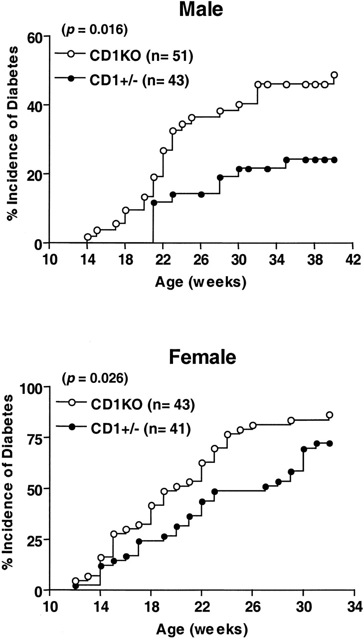
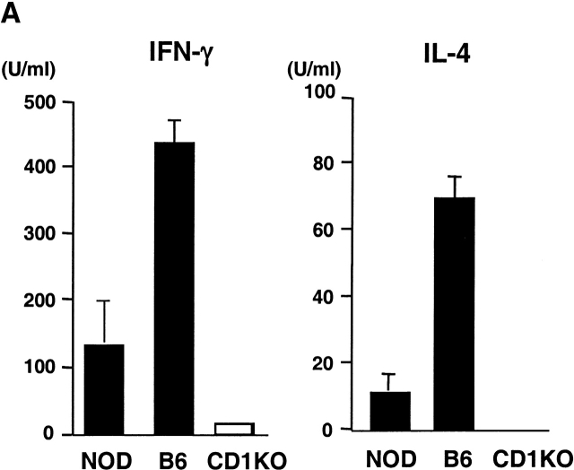
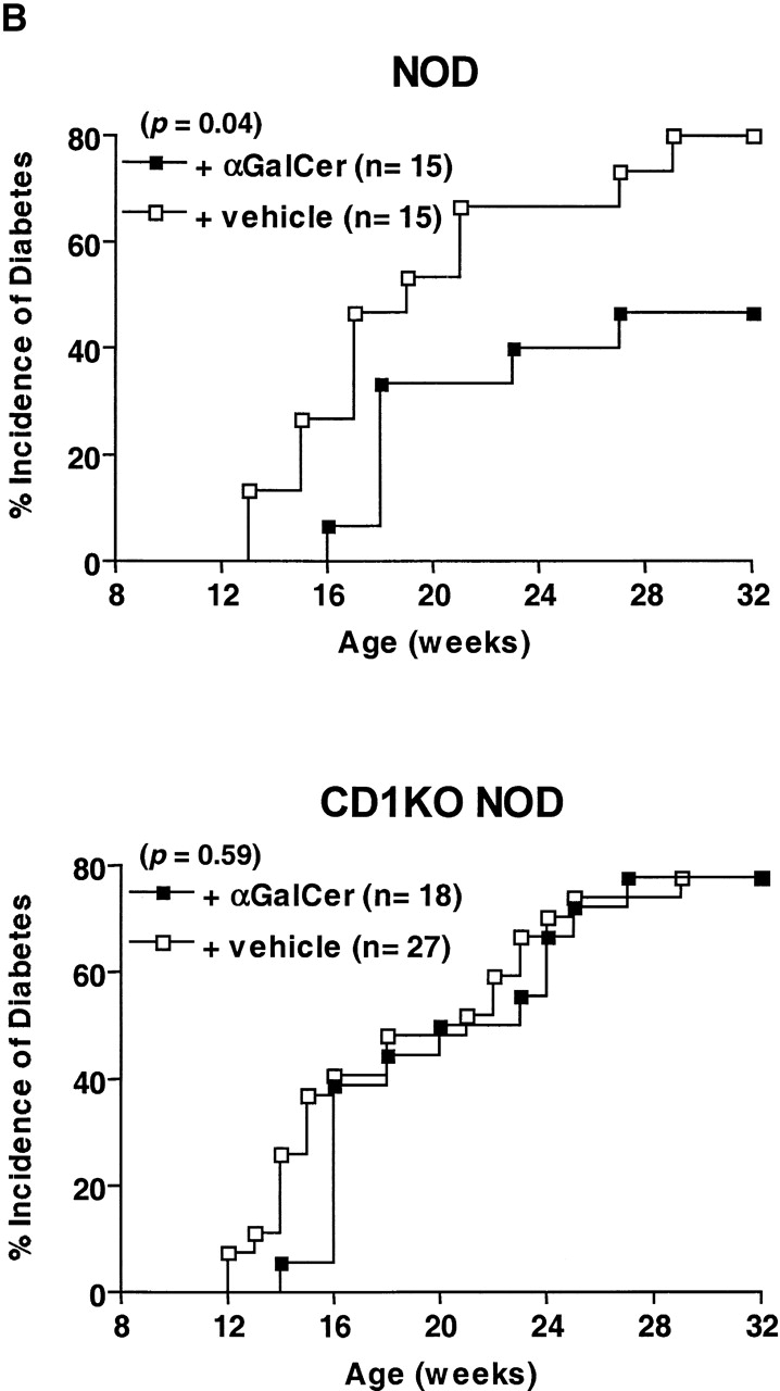
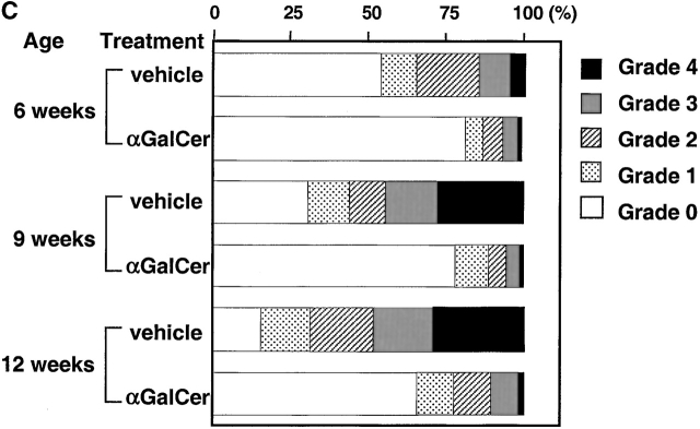
NK T cells are a unique subset of T cells that recognize lipid antigens presented by CD1d. After activation, NK T cells promptly produce large amounts of cytokines, which may modulate the upcoming immune responses. Previous studies have documented an association between decreased numbers of NK T cells and the progression of some autoimmune diseases, suggesting that NK T cells may control the development of autoimmune diseases. To investigate the role of NK T cells in autoimmune diabetes, we crossed CD1 knockout (CD1KO) mutation onto the nonobese diabetic (NOD) genetic background. We found that male CD1KO NOD mice exhibited significantly higher incidence and earlier onset of diabetes compared with the heterozygous controls. The diabetic frequencies in female mice showed a similar pattern; however, the differences were less profound between female CD1KO and control mice. Early treatment of NOD mice with alpha-galactosylceramide, a potent NK T cell activator, reduced the severity of autoimmune diabetes in a CD1-dependent manner. Our results not only suggest a protective role of CD1-restricted NK T cells in autoimmune diabetes but also reveal a causative link between the deficiency of NK T cells and the induction of insulin-dependent diabetes mellitus.


Figures

Figure 1
Figure 1
Increased onset and incidence of diabetes in CD1KO NOD mice. CD1KO mice were backcrossed six to nine generations onto NOD/Lt background and then intercrossed to generate CD1+/− and CD1KO NOD mice. Beginning at 10 wk of age, the urine glucose levels of NOD mice were monitored every 2 wk. Male (top) and female (bottom) mice were considered to have developed diabetes if two consecutive glucosuria levels were >500 mg/dl and blood glucose levels were >250 mg/dl. ○, homozygous CD1-deficient NOD mice; •, CD1 heterozygous NOD littermate mice.
Figure 3
Figure 3
IL-4 and IFN-γ production by splenic T cells from CD1+/− and CD1KO NOD mice. (A) The CD4+-enriched cells (5 × 10⁵ cells per well) from the indicated mice were cultured with immobilized anti-CD3 (10 μg/ml). Supernatants were harvested 48 h later and assayed for IL-4 (top) and IFN-γ (bottom) by ELISA. (B) The ratio of IFN-γ/IL-4 produced by T cells from CD1KO NOD and littermate controls. Data shown represent mean ± SE of four to five mice in each group.
Figure 3
Figure 3
IL-4 and IFN-γ production by splenic T cells from CD1+/− and CD1KO NOD mice. (A) The CD4+-enriched cells (5 × 10⁵ cells per well) from the indicated mice were cultured with immobilized anti-CD3 (10 μg/ml). Supernatants were harvested 48 h later and assayed for IL-4 (top) and IFN-γ (bottom) by ELISA. (B) The ratio of IFN-γ/IL-4 produced by T cells from CD1KO NOD and littermate controls. Data shown represent mean ± SE of four to five mice in each group.
Figure 2
Figure 2
Histopathologic examination of pancreatic insulitis in CD1KO NOD and CD1+/− NOD littermates. Pancreatic tissues were taken at serial time points. After hematoxylin and eosin staining, a minimum of 30 islets from each mouse was examined. The severity of insulitis was determined and graded semiquantitatively using the criteria as described in Materials and Methods. Data shown represent mean of four to five animals in each age group.
Figure 4
Figure 4
α-GalCer protects NOD mice from developing IDDM. (A) In vitro activation of spleen cells from NOD and B6 mice with α-GalCer. Spleen cells (5 × 10⁵ cells per well) from the indicated mice were cultured with 100 ng/ml of α-GalCer. 2 d later, cytokine production in the culture supernatant was measured by ELISA. Spleen cells cultured with vehicle (0.1% DMSO) did not secrete detectable amounts of IFN-γ and IL-4 (<1 U/ml). (B) The effect of α-GalCer on the incidence of diabetes. Female NOD (top) and CD1KO NOD (bottom) mice were injected intraperitoneally twice per week with α-GalCer (▪) or 0.025% polysorbate-20 vehicle (□) as described in Materials and Methods. Starting at 10 wk of age, diabetes was monitored by measurements of urine and blood glucose levels. (C) The effect of α-GalCer on the insulitis of NOD mice. Female NOD mice were treated with α-GalCer or 0.025% polysorbate vehicle starting at 3 wk of age, as described in Materials and Methods. Pancreata were microscopically evaluated for the degree of insulitis as described above. Data shown represent the mean of five animals in each group.
Figure 4
Figure 4
α-GalCer protects NOD mice from developing IDDM. (A) In vitro activation of spleen cells from NOD and B6 mice with α-GalCer. Spleen cells (5 × 10⁵ cells per well) from the indicated mice were cultured with 100 ng/ml of α-GalCer. 2 d later, cytokine production in the culture supernatant was measured by ELISA. Spleen cells cultured with vehicle (0.1% DMSO) did not secrete detectable amounts of IFN-γ and IL-4 (<1 U/ml). (B) The effect of α-GalCer on the incidence of diabetes. Female NOD (top) and CD1KO NOD (bottom) mice were injected intraperitoneally twice per week with α-GalCer (▪) or 0.025% polysorbate-20 vehicle (□) as described in Materials and Methods. Starting at 10 wk of age, diabetes was monitored by measurements of urine and blood glucose levels. (C) The effect of α-GalCer on the insulitis of NOD mice. Female NOD mice were treated with α-GalCer or 0.025% polysorbate vehicle starting at 3 wk of age, as described in Materials and Methods. Pancreata were microscopically evaluated for the degree of insulitis as described above. Data shown represent the mean of five animals in each group.

References

    1. Kikutani H., Makino S. The murine autoimmune diabetes modelNOD and related strains. Adv. Immunol. 1992;51:285–322. - PubMed
    1. Bendelac A., Carnaud C., Boitard C., Bach J.F. Syngeneic transfer of autoimmune diabetes from diabetic NOD mice to healthy neonates. Requirement for both L3T4+ and Lyt-2+ T cells. J. Exp. Med. 1987;166:823–832. - PMC - PubMed
    1. Miller B.J., Appel M.C., O'Neil J.J., Wicker L.S. Both the Lyt-2+ and L3T4+ T cell subsets are required for the transfer of diabetes in nonobese diabetic mice. J. Immunol. 1988;140:52–58. - PubMed
    1. Yagi H., Matsumoto M., Kunimoto K., Kawaguchi J., Makino S., Harada M. Analysis of the roles of CD4+ and CD8+ T cells in autoimmune diabetes of NOD mice using transfer to NOD athymic nude mice. Eur. J. Immunol. 1992;22:2387–2393. - PubMed
    1. Katz J.D., Benoist C., Mathis D. T helper cell subsets in insulin-dependent diabetes. Science. 1995;268:1185–1188. - PubMed

Publication types

MeSH terms