. 2001 Jul;128(13):2497-508.
doi: 10.1242/dev.128.13.2497.

Math5 is required for retinal ganglion cell and optic nerve formation

Affiliations

Math5 is required for retinal ganglion cell and optic nerve formation

N L Brown et al. Development. 2001 Jul.

Abstract

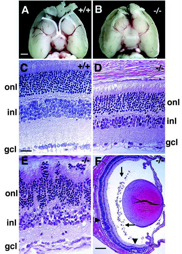
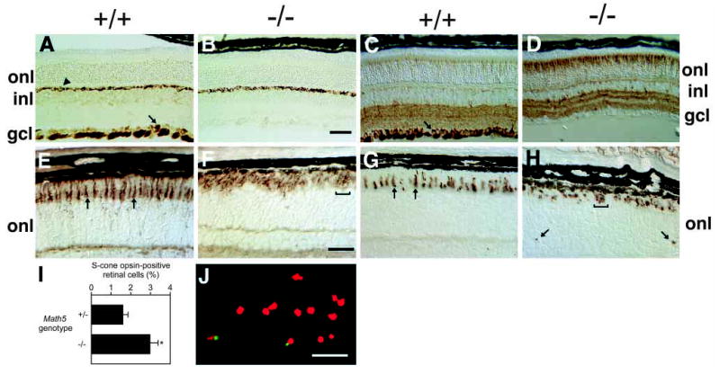
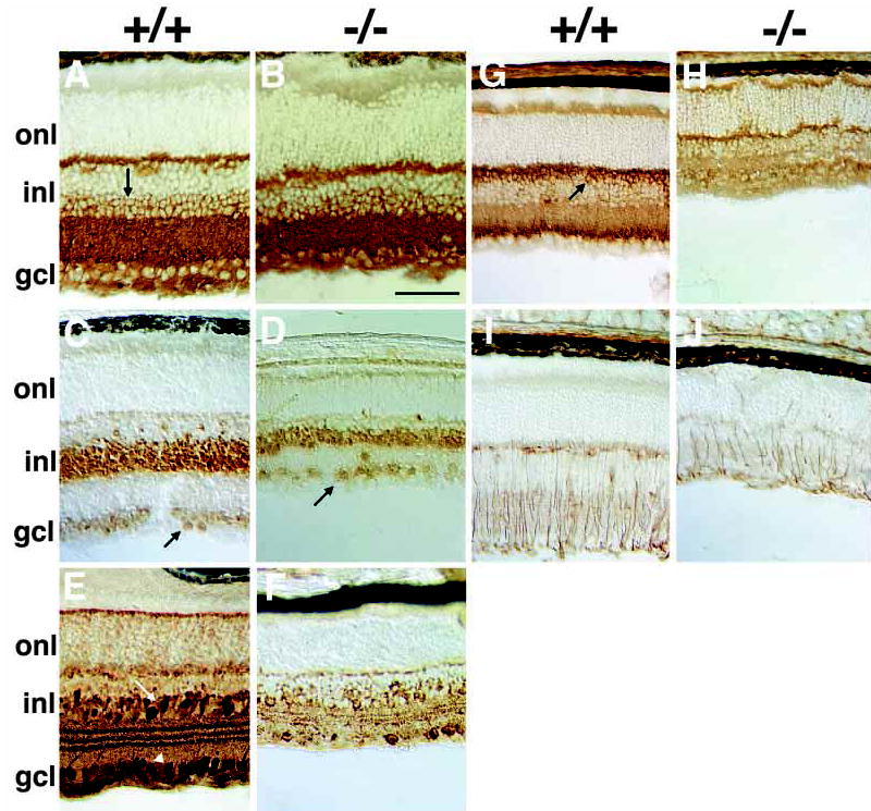
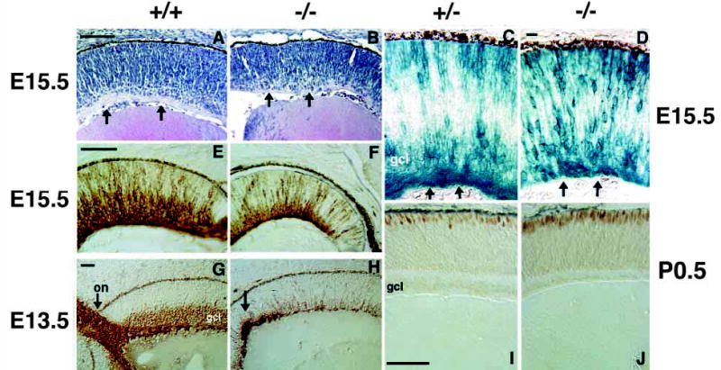
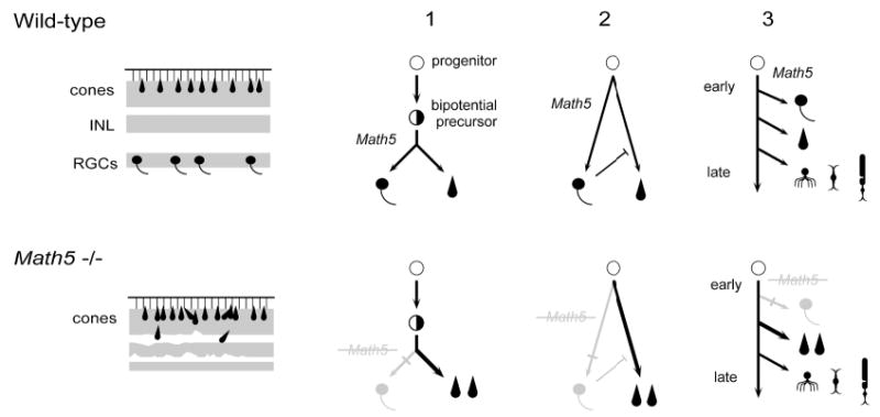
The vertebrate retina contains seven major neuronal and glial cell types in an interconnected network that collects, processes and sends visual signals through the optic nerve to the brain. Retinal neuron differentiation is thought to require both intrinsic and extrinsic factors, yet few intrinsic gene products have been identified that direct this process. Math5 (Atoh7) encodes a basic helix-loop-helix (bHLH) transcription factor that is specifically expressed by mouse retinal progenitors. Math5 is highly homologous to atonal, which is critically required for R8 neuron formation during Drosophila eye development. Like R8 cells in the fly eye, retinal ganglion cells (RGCs) are the first neurons in the vertebrate eye. Here we show that Math5 mutant mice are fully viable, yet lack RGCs and optic nerves. Thus, two evolutionarily diverse eye types require atonal gene family function for the earliest stages of retinal neuron formation. At the same time, the abundance of cone photoreceptors is significantly increased in Math5(-/-) retinae, suggesting a binary change in cell fate from RGCs to cones. A small number of nascent RGCs are detected during embryogenesis, but these fail to develop further, suggesting that committed RGCs may also require Math5 function.


Figures

Fig. 1
Fig. 1
Targeted disruption of Math5. (A) Homologous recombination between 2.3 kb and 4 kb vector arms resulted in replacement of the bHLH domain (contained within the SmaI fragment) with a PGK-neo cassette and insertion of cytoplasmic β-gal near the N terminus of Math5. Positions of the single-copy probe and PCR primers are indicated. Primers A+A′ (5′ arm) and B+B′ (3′ arm) were used for long-range PCR. (B) BamHI Southern analysis of littermate tail DNA obtained from an F2 cross. (C) Internal PCR primers (D+D′ for wild-type, C+C′ for mutant) amplify allele-specific products from weanling tail DNA. (D) RT-PCR products (F+F′) from E15.5 total eye RNA show the absence of Math5 mRNA and a great reduction of Brn3b mRNA in the Math5−/− mutant, in comparison to β-actin. B, BamHI; E, EcoRI; H, HindIII; S, SmaI.
Fig. 2
Fig. 2
Embryonic β-gal expression in Math5+/− eyes. (A,B) E11.5 β-gal expression in the developing optic cup. B shows a horizontal section through the whole-mount embryo in A. The overall pattern is identical to endogenous Math5 mRNA (see Fig. 2 of Brown et al., 1998). β-gal staining begins in the dorsal central cup. A small number of positive cells are also visible in the diencephalon near the base of the optic stalk. (C,D) E15.5 retinal β-gal expression. β-gal is present in progenitors and differentiating RGCs. D shows a higher magnification. E. In situ hybridization at E15.5 showing Math5 mRNA expression solely in retinal progenitors. (F) β-gal expression in the optic nerve at day E17.5 (arrow). (G) β-gal-positive cells with characteristic cone morphology in the photoreceptor layer of a heterozygote at P21. Bars are 1 mm in A; 200 μm in B; 100 μm in D; 500 μm in F; and 20 μm in G.
Fig. 3
Fig. 3
Ocular abnormalities in adult Math5−/− mice. (A,B) Ventral view of Math5+/+ and Math5−/− P21 brains, with rostral ends pointing upwards. Olfactory bulbs have been removed. Both optic nerves and the optic chiasm are absent in the Math5−/− mouse. (C–F) Hematoxylin and Eosin-stained transverse sections of P21 Math5+/+ (C) and Math5−/− (D–F) eyes. Mutant retinae have regions of normal (D,F) and disrupted laminar structure (E and arrowheads in F). However, even in areas with largely normal structure, cells and axons are missing from the INL, IPL and GCL (D,E). Ectopic blood vessels are present in the vitreal space of mutant eyes (arrows in F). gcl, ganglion cell layer; inl, inner nuclear layer; onl, outer nuclear layer. Bars are 1.5 mm in A; 20 μm in C; and 200 μm in F.
Fig. 4
Fig. 4
RGCs are absent and cone photoreceptors are increased in Math5−/− mice. (A,B) Neurofilament (160 kDa) is normally expressed by RGCs (arrow in A) and horizontal neurons (arrowhead in A). This marker highlights the loss of RGCs and their axon fibers in Math5 mutants (B). Horizontal cells are unaffected. (C,D) Anti-β-tubulin (TUJ1) labeling shows the absence of RGCs and an excess of cone photoreceptors in Math5−/− retinae. RGCs are strongly labeled in the wild-type section (arrow). (E,F) Peanut agglutinin (PNA) stains all cone photoreceptors. Individual cones are visible in the wild-type (arrows in E) but the density of cones is greatly increased in the Math5−/− mice (F). (G,H) The abundance of S-cones is also increased in Math5 mutants. The arrows in G indicate normal S-cone outer segments. Mutant cones are irregularly arranged in the outer retina (brackets in F and H). In some mutant sections, S-cone outer segments were observed within the ONL (arrows in H), OPL or INL. (I) Histogram comparing the abundance of S-cones in Math5+/− and Math5−/− mice as a percentage of total retinal cells. The bars show mean values and standard errors for these two genotypes. The increase in cones in Math5−/− mice is statistically significant (t s[∞] = 2.74, P <0.01). (J) Dissociated retinal cells labeled with S-cone opsin antisera (green) and propidium iodide (red). All retinal sections (A–H) are from P21 mice. gcl, ganglion cell layer; inl, inner nuclear layer; onl, outer nuclear layer. Bars are 100 μm in B; 30 μm in F; 50 μm in J.
Fig. 5
Fig. 5
Other retinal neuron defects in Math5−/− mice. (A,B) Anti-syntaxin (HPC-1) immunostaining shows that amacrines in vitread INL (arrow) and displaced amacrines in the GCL are grossly unaffected. (C,D) Pax6 is similarly expressed by INL and displaced amacrine cells of wild-type (C) and Math5 mutant mice (D). (E,F) Calretinin-positive A2 amacrines in the INL (arrow) and GCL (arrowhead) are reduced in the Math5 mutant (F) compared to wild-type (E). (G,H) Rod bipolar cells labeled with anti-protein kinase C have cell bodies in the sclerad INL (arrow) and synapse with photoreceptors in the OPL and A2 amacrines in the IPL. These neurons are reduced in Math5−/− retinae and lack well organized synaptic termini in the IPL. (I,J) Anti-vimentin staining shows radial fibers of Müller glia extending across Math5+/+ retinal laminae. The abundance of Müller glia is reduced in Math5−/− eyes. Retinal sections in A–D are from P14 mice and those in E–J are from P21. gcl, ganglion cell layer; inl, inner nuclear layer; onl, outer nuclear layer. The bar is 100 μm in B.
Fig. 6
Fig. 6
Early stages of RGC and cone formation are abnormal in Math5−/− retinae. Micrographs of retinal sections from E15.5 (A–F) and E13.5 (G,H) embryos and P0.5 newborn mice (I,J). (A,B) Hematoxylin and Eosin-stained sections of wild-type (A) and Math5−/− (B) retinae. Mutant retinae are thinner in the radial dimension. Arrows in both panels indicate the position of eosinophilic RGC axon fibers, which are apparent in A but largely absent in B. (C,D) β-gal expression in Math5+/− and Math5−/− retinal sections. Arrows indicate the RGC axon fibers in heterozygotes that are greatly reduced in the mutant. Math5−/− β-gal-expressing cells are distributed across the retinal thickness (top to bottom of D). (E,F) Anti-β-tubulin (TUJ1) immunostaining of E15.5 retinae. A well formed GCL is apparent in the wild-type (E), but is greatly diminished in the Math5−/− retina (F). (G,H) Neurofilament (160 kDa) immunostaining highlights the failure of Math5−/− retinae to form a significant number of RGCs at an early stage. Arrows indicate a normal optic nerve in wild-type (G) and abnormal axons in the Math5 mutant that do not exit the eye (H). (I,J) Recoverin immunostaining demonstrates an early increase in the density of cone photoreceptors in Math5−/− mice (J). on, optic nerve; gcl, ganglion cell layer. Bars are 100 μm in A,E,I; 20 μm in D; and 50 μm in G.
Fig. 7
Fig. 7
Binary cell fate switch between RGCs and cones in Math5−/− mice. On the left, diagrams summarize wild-type (upper) and mutant (lower) retinal phenotypes. RGCs are missing and cones are increased in the mutant. The nuclear layers are indicated by shading. The inner nuclear (INL) and plexiform layers are significantly thinner in the mutant and lamination is disrupted in places. On the right, diagrams depict three models that explain the shift from RGCs to cones, which are outlined in the text. In addition to the multipotent progenitor and hypothetical bipotential precursor cells, five differentiated retinal cell types are represented (RGCs, cones, amacrines, bipolars, and rods).

References

    1. Altshuler D, Cepko C. A temporally regulated, diffusible activity is required for rod photoreceptor development in vitro. Development. 1992;114:947–957. - PubMed
    1. Altshuler, D. M., Turner, D. L. and Cepko, C. L. (1991). Specification of cell type in the vertebrate retina. In Cell Lineage and Cell fate in Visual System Development (ed. D. M.-K. Lam and C. J. Shatz), pp. 37–58. Cambridge, MA: MIT Press.
    1. Applebury ML, Antoch MP, Baxter LC, Chun LL, Falk JD, Farhangfar F, Kage K, Krzystolik MG, Lyass LA, Robbins JT. The murine cone photoreceptor: a single cone type expresses both S and M opsins with retinal spatial patterning. Neuron. 2000;27:513–523. - PubMed
    1. Austin CP, Feldman DE, Ida JA, Cepko CL. Vertebrate retinal ganglion cells are selected from competent progenitors by the action of Notch. Development. 1995;121:3637–3650. - PubMed
    1. Ballesta J, Terenghi G, Thibault J, Polak JM. Putative dopamine-containing cells in the retina of seven species demonstrated by tyrosine hydroxylase immunocytochemistry. Neuroscience. 1984;12:1147–1156. - PubMed

Publication types

MeSH terms

Substances