. 2001 Aug 14;98(17):9784-9.
doi: 10.1073/pnas.171269898. Epub 2001 Aug 7.

Nephrocystin interacts with Pyk2, p130(Cas), and tensin and triggers phosphorylation of Pyk2

Affiliations

Nephrocystin interacts with Pyk2, p130(Cas), and tensin and triggers phosphorylation of Pyk2

T Benzing et al. Proc Natl Acad Sci U S A. .

Abstract

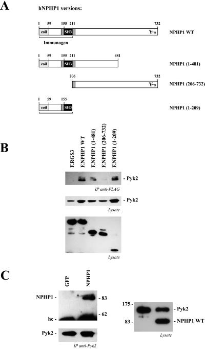
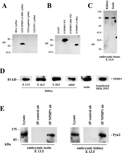
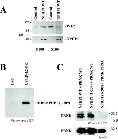
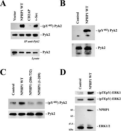
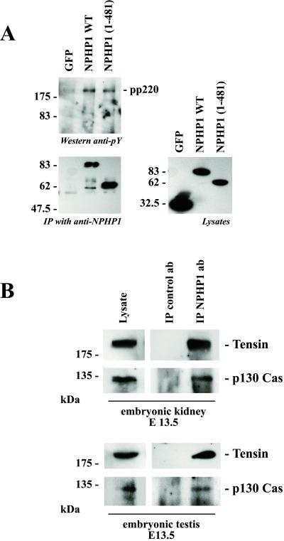
Juvenile nephronophthisis type 1 is caused by mutations of NPHP1, the gene encoding for nephrocystin. The function of nephrocystin is presently unknown, but the presence of a Src homology 3 domain and its recently described interaction with p130(Cas) suggest that nephrocystin is part of the focal adhesion signaling complex. We generated a nephrocystin-specific antiserum and analyzed the interaction of native nephrocystin with endogenous proteins. Immunoprecipitation of nephrocystin revealed that nephrocystin forms protein complexes with p130(Cas), proline-rich tyrosine kinase 2 (Pyk2), and tensin, indicating that these proteins participate in a common signaling pathway. Expression of nephrocystin resulted in phosphorylation of Pyk2 on tyrosine 402 as well as activation of downstream mitogen-activated protein kinases, such as ERK1 and ERK2. Our findings suggest that nephrocystin helps to recruit Pyk2 to cell matrix adhesions, thereby initiating phosphorylation of Pyk2 and Pyk2-dependent signaling. A lack of functional nephrocystin may compromise Pyk2 signaling in a subset of renal epithelial cells.


Figures

Figure 1
Figure 1
Nephrocystin interacts with Pyk2. (A) Several Flag-tagged nephrocystin truncations were generated to analyze the interaction with Pyk2. Nephrocystin contains several putative protein–protein interaction motifs, including an amino-terminal coiled-coil domain (coil) and an SH3 domain. (B) Nephrocystin precipitates Pyk2. HA-tagged Pyk2 and Flag-tagged nephrocystin constructs were expressed in HEK 293T cells and precipitated with a Flag-specific antibody. Western blot analysis was performed with an HA-specific antiserum (Top and Middle). Pyk2 coprecipitates with nephrocystin but not with the control protein RGS3 (Top). The interaction requires the N-terminal region of nephrocystin, containing the SH3 domain. (Middle) Expression levels of HA.Pyk2 are shown. (Bottom) Expression levels of Flag-tagged RGS3 and nephrocystin constructs in cell lysates are shown. (C) Pyk2 precipitates nephrocystin but not the control protein GFP (Left Upper); the Pyk2 precipitates are shown (Left Lower). (Right) The expressions of Flag-tagged nephrocystin and HA-tagged Pyk2 in the cellular lysates are shown.
Figure 2
Figure 2
Interaction between endogenous nephrocystin and Pyk2. (A and B) To generate a nephrocystin-specific antiserum, rabbits were immunized with a recombinant MBP fusion protein, containing the amino-terminal 209 aa of nephrocystin. Affinity-purified antiserum specifically recognized recombinant nephrocystin fused to GST and nephrocystin expressed in HEK 293T cells, but failed to bind recombinant control proteins or a nephrocystin version lacking the region used for immunization. (C and D) The antiserum recognizes mouse nephrocystin in endogenous tissues. To test whether mouse nephrocystin can be detected in embryonic tissues, mouse embryonic kidneys and testes were lysed at different developmental stages, and the solubilized proteins were separated on a 10% SDS/PAGE. Western blot analysis revealed that the antiserum raised against human nephrocystin detects mouse nephrocystin. (E) Nephrocystin interacts with Pyk2 in embryonic tissues. The nephrocystin antiserum was used to examine the endogenous interaction between Pyk2 and nephrocystin in the embryonic mouse kidney and testis. Embryonic kidneys (Right) and embryonic testes (Left) were collected and homogenized. The lysates were immunoprecipitated with a control antiserum or the nephrocystin-specific antiserum. Western blot analysis was performed with a Pyk2-specific antiserum.
Figure 3
Figure 3
Subcellular localization of Pyk2 and direct interaction between nephrocystin and the proline-rich (PR) region of Pyk2. (A) Nephrocystin modulates the subcellular localization of Pyk2. The tubular epithelial cell line IMCD was infected with a retrovirus, directing the expression of nephrocystin, or a control retrovirus. Nephrocystin increased the membrane-associated fraction (P100) and decreased the cytoplasmic fraction (S100) of Pyk2. (B) GST and GST.Pyk2-PR were immobilized on glutathione-Sepharose beads and incubated with MBP.NPHP1 (1). NPHP1 binds to GST.Pyk2-PR but not to GST. (C) Coimmunoprecipitation of PRNK, a naturally occurring splice variant of Pyk2, with full-length nephrocystin (NPHP1 WT) or the C-terminally truncated version [NPHP1 (1)]. (Upper) Mutation of the proline residue 859 in Pyk2 abrogates the interaction with nephrocystin. (Lower) Expression of wild-type and mutated PRNK is shown.
Figure 4
Figure 4
Nephrocystin triggers phosphorylation of tyrosine 402 of Pyk2. (A) HEK 293T cells were transfected with HA-tagged Pyk2 in combination with vector control, nephrocystin, the SH3 domain-containing adapter protein CD2AP, or the positive control v-Src. HA.Pyk2 was immunoprecipitated from serum-starved cells with anti-HA antibody; precipitates were probed with an antiserum detecting phosphotyrosine 402 of Pyk2 (Top) or with an antiserum revealing the total amount of Pyk2 (Middle). (Bottom) The expression of HA.Pyk2 in the lysates. (B) Pyk2 is phosphorylated on tyrosine 402 in nephrocystin-expressing IMCD cells. IMCD cells were infected with either a control or a nephrocystin-expressing retrovirus. Nephrocystin induces tyrosine 402 phosphorylation of endogenous Pyk2 in IMCD cells (Upper) but does not influence the expression levels of Pyk2 (Lower). (C) Full-length nephrocystin is required to trigger phosphorylation of tyrosine 402 of Pyk2. Retroviral vectors, expressing nephrocystin truncations, were constructed to infect IMCD cells. Both the amino-terminal deletion [NPHP1 (206)] and the carboxyl-terminal deletion [NPHP1 (1)] of nephrocystin failed to mediate tyrosine 402 phosphorylation of Pyk2 (Upper). (Lower) The levels of Pyk2 expression are shown. (D) Nephrocystin activates ERK1/2. Dually phosphorylated ERK1 and ERK2 was visualized in IMCD cells, transduced with control vector or a retroviral vector encoding for nephrocystin, using a phosphospecific antiserum (Upper). The blots were reprobed with an antiserum detecting total amounts of ERK1 and ERK2 and with the nephrocystin-specific antiserum.
Figure 5
Figure 5
Nephrocystin associates with p130Cas and tensin. (A) To identify nephrocystin-interacting proteins, HEK 293T cells were transfected with the control protein GFP, nephrocystin, or the nephrocystin truncation NPHP1 (1). After immunoprecipitation of nephrocystin, immunoprecipitates were separated on a 10% SDS/PAGE and stained with the monoclonal antiphosphotyrosine antibody 4G10 (Left Upper), or anti-Flag antibody (Left Lower). A tyrosine-phosphorylated protein with an approximate molecular mass of 220 kDa (pp220) was detected in the nephrocystin precipitates. Expression levels of Flag-tagged GFP and nephrocystin truncations in the lysates are demonstrated (Right). HEK 293T cells lack endogenous Pyk2, and consequently Pyk2 was not detectable in the 4G10-labeled precipitates. (B) Coprecipitation of tensin and p130Cas with native nephrocystin from embryonic tissues. Embryonic kidneys (Upper) and embryonic testes (Lower) were collected and homogenized. The lysates were immunoprecipitated with a control or nephrocystin-specific antiserum and separated by 10% SDS/PAGE. Tensin and p130Cas were detected in the nephrocystin-containing precipitates using tensin or p130Cas-specific antibodies.

References

    1. Hildebrandt F, Otto E. J Am Soc Nephrol. 2000;11:1753–1761. - PubMed
    1. Waldherr R, Lennert T, Weber H P, Fodisch H J, Scharer K. Virchows Arch Pathol Anat Physiol Klin Med. 1982;394:235–254. - PubMed
    1. Hildebrandt F, Otto E, Rensing C, Nothwang H G, Vollmer M, Adolphs J, Hanusch H, Brandis M. Nat Genet. 1997;17:149–153. - PubMed
    1. Saunier S, Calado J, Heilig R, Silbermann F, Benessy F, Morin G, Konrad M, Broyer M, Gubler M C, Weissenbach J, Antignac C. Hum Mol Genet. 1997;6:2317–2323. - PubMed
    1. Sherman F E, Studnicki F M, Fetterman G. Am J Clin Pathol. 1971;55:391–400. - PubMed

Publication types

MeSH terms