. 2001 Aug 14;98(17):9666-70.
doi: 10.1073/pnas.171301998. Epub 2001 Aug 7.

p70S6 kinase signals cell survival as well as growth, inactivating the pro-apoptotic molecule BAD

Affiliations

p70S6 kinase signals cell survival as well as growth, inactivating the pro-apoptotic molecule BAD

H Harada et al. Proc Natl Acad Sci U S A. .

Abstract

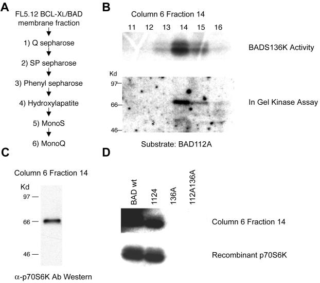
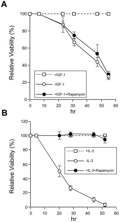
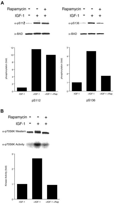
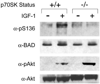
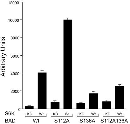
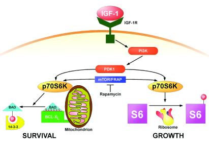
Cytokines often deliver simultaneous, yet distinct, cell growth and cell survival signals. The 70-kDa ribosomal protein S6 kinase (p70S6K) is known to regulate cell growth by inducing protein synthesis components. We purified membrane-based p70S6K as a kinase responsible for site-specific phosphorylation of BAD, which inactivates this proapoptotic molecule. Rapamycin inhibited mitochondrial-based p70S6K, which prevented phosphorylation of Ser-136 on BAD and blocked cell survival induced by insulin-like growth factor 1 (IGF-1). Moreover, IGF-1-induced phosphorylation of BAD Ser-136 was abolished in p70S6K-deficient cells. Thus, p70S6K is itself a dual pathway kinase, signaling cell survival as well as growth through differential substrates which include mitochondrial BAD and the ribosomal subunit S6, respectively.


Figures

Figure 1
Figure 1
Purification of the BAD S136 specific kinase. (A) The purification scheme used for a BAD S136-specific kinase. (B Upper) Fractions 11–16 from the Mono Q column were assayed for BAD kinase activity with GST-BAD S112A as a substrate. (Lower) In-gel kinase assay was performed by using the same fractions as above with GST-BAD S112A as a substrate. (C) A Western blot of fraction 14 from the Mono Q column using an anti-p70S6K antibody. (D) Purified BAD kinase (Mono Q, fraction 14) was tested for its ability to phosphorylate GST-BAD wild-type (BAD wt), and GST-BAD whose S112, S136, or both were changed to Ala (112A, 136A, and 112A136A). Recombinant p70S6K (Upstate Biotechnology, Lake Placid, NY) was used to show S136 specificity of BAD phosphorylation in vitro.
Figure 2
Figure 2
Inhibition of p70S6K activation induced by IGF-1 enhances cell death. FL5.12 BCL-XL/BAD S112A cells were deprived of IL-3 with or without rapamycin (20 ng/ml, Calbiochem) to initiate a death signal, whereas IGF-1 (50 ng/ml, Life Technologies) or IL-3 (10 ng/ml, Genzyme) was added back at 4 h before irreversible damage occurred. At each time point, viability was assessed by trypan blue exclusion. The IGF-1- or IL-3-dependent survival is compared with and without rapamycin. Data shown are representative of three independent experiments.
Figure 3
Figure 3
Rapamycin specifically inhibits IGF-1-induced BAD S136 phosphorylation and p70S6K activity in the mitochondrial fraction. (A) Survival factors were withdrawn from FL5.12 BCL-XL/BAD S112A or FL5.12 BCL-XL/BAD S136A cells for 4 h (−IGF-1), followed by addition of IGF-1 (50 ng/ml) for 15 min (+IGF-1). Rapamycin (20 ng/ml) was added when factors were withdrawn. Western blot analyses were done with antibodies that recognize phosphorylated forms of BAD at S112 or S136 (19, 22). The filters were reprobed with an antibody that recognizes BAD regardless of its phosphorylation state. Data shown are representative of two independent experiments. Histograms present the fold increase in intensity of the phosphorylated band relative to total BAD, in which − IGF-1 is set at 1.0. (B) FL5.12 BCL-XL/BAD S112A cells were treated as in A. The mitochondrial heavy membrane fractions were analyzed by Western blot with an anti-p70S6K Ab (Upper), or immunoprecipitated with an anti-p70S6K Ab, followed by an in vitro kinase assay using GST-S6 as substrate. Data shown are representative of two independent experiments. Histogram presents the fold increase in intensity of the phosphorylated S6.
Figure 4
Figure 4
IGF-1-induced phosphorylation of BAD S136 is abolished in p70S6K-deficient cells. Serum was withdrawn from p70S6K+/+ or p70S6K−/− ES cells for 4 h (−IGF-1), followed by addition of IGF-1 (50 ng/ml) for 15 min (+IGF-1). Whole-cell extracts were immunoprecipitated with an anti-BAD antibody, followed by Western blot analyses with a phospho-S136-specific anti-BAD antibody or an antibody that recognizes BAD regardless of its phosphorylation state. Western blot analyses were also done by using an antibody that recognizes the phosphorylated form of AKT or an antibody that recognizes AKT regardless of its phosphorylation state. Data shown are representative of two independent experiments.
Figure 5
Figure 5
p70S6K suppresses BAD-mediated death in a S136-dependent manner. Rat-1a cells were transiently cotransfected with a luciferase reporter together with constructs expressing either BAD wild-type (Wt), S112A, S136A, or S112AS136A together with constructs expressing either p70S6K Wt or a kinase-dead (KD) mutant (Lys-100 → Gln) that does not bind ATP. Twenty-four hours after transfection, serum was withdrawn for 16 h. Luciferase activity (ordinate, arbitrary units) has been shown to represent the viability of the cells defined (9, 27), and this assay is representative of three independent experiments.
Figure 6
Figure 6
p70S6K as a dual pathway kinase. A schematic diagram of survival and growth substrates of S6K.IGF-1R, IGF-1 receptor; PI3K, phosphatidylinositol 3-kinase; PDK-1, 3-phosphoinositide-dependent protein kinase 1.

References

    1. Conlon I, Raff M. Cell. 1999;96:235–244. - PubMed
    1. Harrington E A, Bennett M R, Fanidi A, Evan G I. EMBO J. 1994;13:3286–3295. - PMC - PubMed
    1. Peruzzi F, Prisco M, Dews M, Salomoni P, Grassilli E, Romano G, Calabretta B, Baserga R. Mol Cell Biol. 1999;19:7203–7215. - PMC - PubMed
    1. Yao R, Cooper G M. Science. 1995;267:2003–2006. - PubMed
    1. Kinoshita T, Yokota T, Arai K, Miyajima A. EMBO J. 1995;14:266–275. - PMC - PubMed

Publication types

MeSH terms