. 2001 Aug 14;98(17):9847-52.
doi: 10.1073/pnas.171319698. Epub 2001 Aug 7.

Hepatitis C virus replication is directly inhibited by IFN-alpha in a full-length binary expression system

Affiliations

Hepatitis C virus replication is directly inhibited by IFN-alpha in a full-length binary expression system

R T Chung et al. Proc Natl Acad Sci U S A. .

Abstract

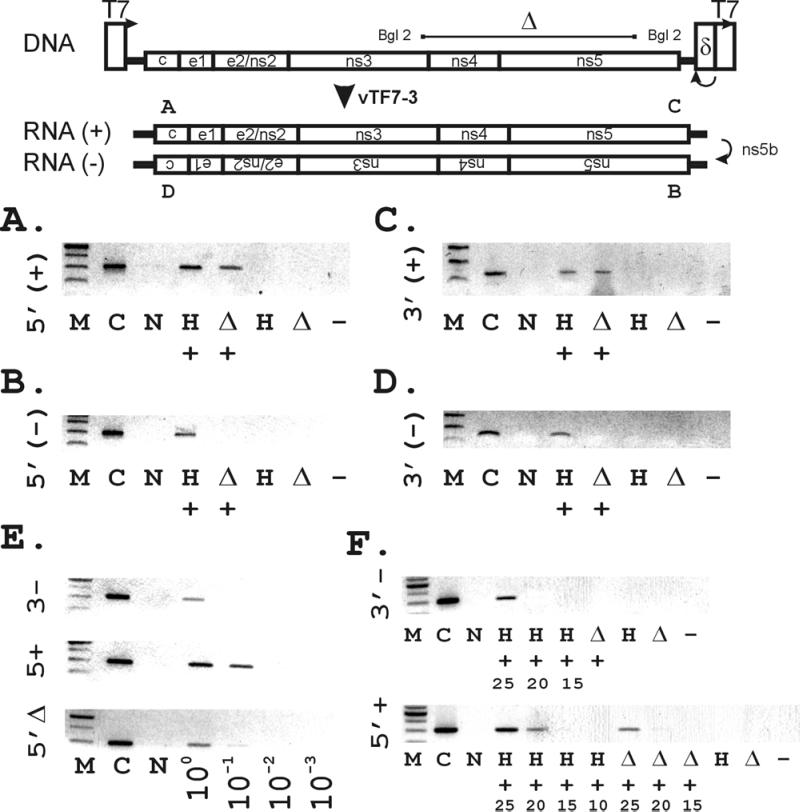
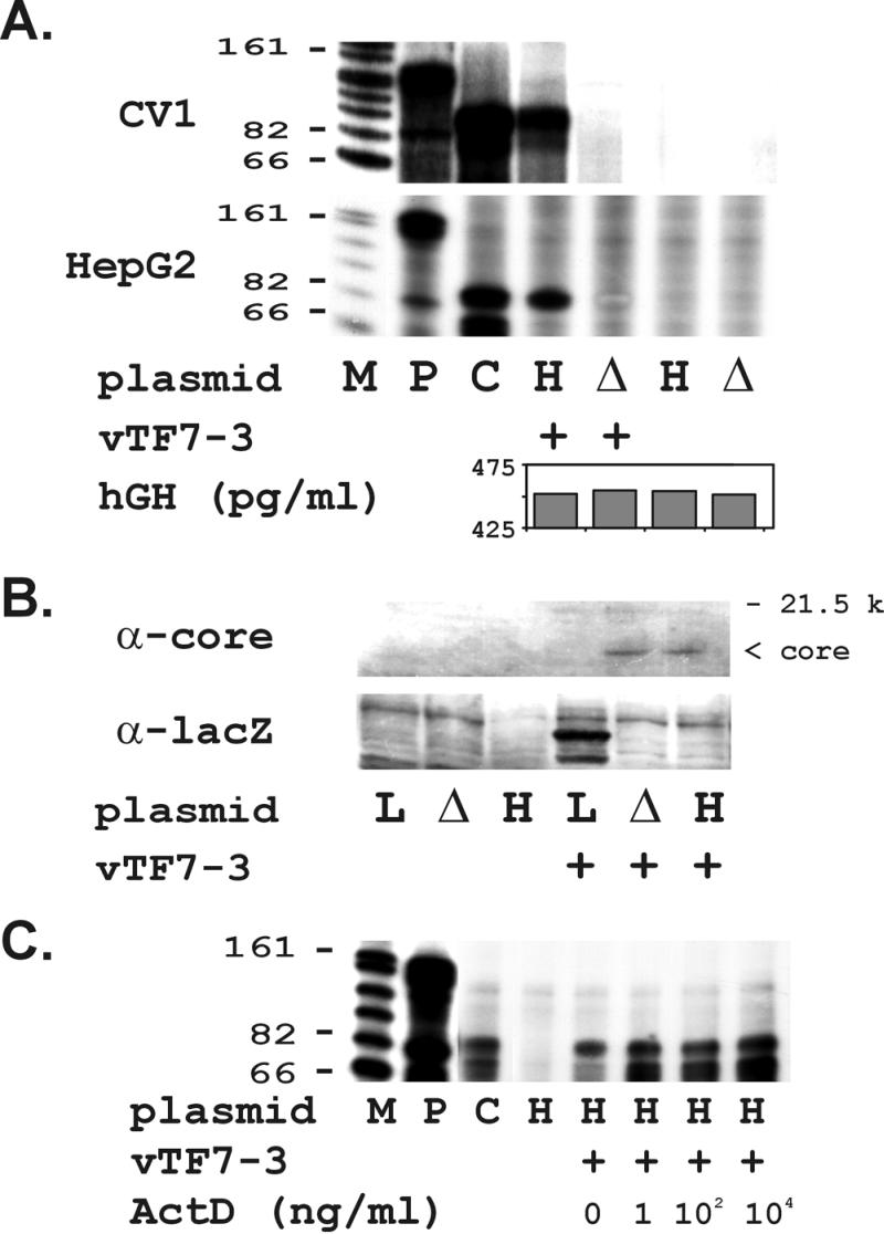
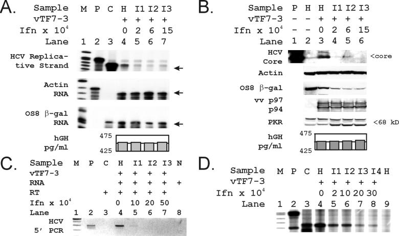
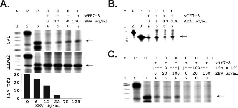
Hepatitis C virus (HCV) is a leading cause of chronic hepatitis, cirrhosis, and hepatocellular carcinoma. The absence of culture systems permissive for HCV replication has presented a major bottleneck to antiviral development. We sought to recapitulate the early steps in the life cycle of HCV by means of DNA-based expression of viral genomic sequences. Here we report expression of replicating HCV RNA by using a, to our knowledge, novel binary expression system in which cells were transfected with a T7 polymerase-driven full-length HCV cDNA plasmid containing a cis-acting hepatitis Delta ribozyme to control 3' cleavage, and infected with vaccinia-T7 polymerase. HCV genomic and replicative strand synthesis, in addition to protein synthesis, was detectable and depended on full-length HCV sequences. Moreover, the system was capable of generating HCV RNA quasispecies, consistent with the action of the low-fidelity HCV NS5B RNA polymerase. IFN-alpha, but not ribavirin, directly inhibited the viral replicative cycle in these cells, identifying the virus itself and not solely the immune system as a direct target of IFN action. The availability of a cell-based test for viral replication will facilitate screening of inhibitory compounds, analysis of IFN-resistance mechanisms, and analysis of virus-host cell interactions.


Figures

Figure 1
Figure 1
(Upper) Construction of vectors used for the binary HCV replication system. The wild-type full-length HCV sequence (pCV-H77) was adapted at its 5′ terminus with the T7 promoter, and at its 3′ terminus with the HDV ribozyme, and the T7 terminator sequence as described in Methods) to generate pT7-flHCV-Rz. A BglII-BglII fragment was excised to create the mutant pT7-HCVΔBglII-Rz as a negative control. (Lower) Synthesis of (+) and (−) strand HCV RNA by RT-PCR depends on the presence of both HCV sequences and T7. Strand-specific RT-PCR was performed as described in Methods, using primers corresponding to the genomic 5′ (A) and 3′ (C) termini and the antigenomic 5′ (B) and 3′ termini (D). (Lanes are designated as follows: M, 100-bp marker; C, pT7flHCVRz DNA-positive control; N, No RNA; H, pT7flHCVRz transfected; Δ, pT7-HCVΔBglII-Rz; +, vaccinia virus vTF7–3 added to supply T7 RNA polymerase; −, no reverse transcriptase added to vTF7–3/pT7flHCVRz RNA.) (E). Limiting dilution analysis of extracted RNAs by using PCR specific for the 5′ full-length genomic (5′+), 3′ full-length antigenomic (3′−), and 5′ BglII genomic strands, using the same sets of primers for each amplification. The remainder of lanes correspond to pT7flHCVRz transfected at the indicated dilutions of RNA. (F) Assessment of (5′+) and (3′−) full-length HCV RNA levels by examination of the cycle dependence of amplified products after strand-specific PCR. Numbers indicated represent cycle number. Lanes without numbers represent PCR products after 25 cycles of amplification.
Figure 2
Figure 2
(A) Synthesis of (−) strand HCV RNA depends on full-length HCV sequences and T7 polymerase and can be supported in multiple mammalian cell lines. RPA for 3′(−) strand HCV RNA detection in transfected CV-1 cells was performed by using a radiolabeled HCV 5′ sense probe as described in Methods. Equivalent results were observed in CV-1 (Upper) and HepG2 (Lower) cell lines. (Lanes are designated as follows: M, RNA marker with sizes indicated; P, free probe; C, H77DNA positive control; H, transfected with pT7-flHCV-Rz; Δ, transfected with pT7-flHCVΔBglII-Rz; +, vaccinia vector vTF7–3 added.) hGH ELISA assessed transfection efficiency for lysates cotransfected with pSVtkhGH. (B) Full-length and truncated HCV RNA transcripts are translated into HCV proteins. (Upper) Detection of HCV core protein synthesis in CV1 cells by using polyclonal human antisera reactive by EIA-2. (Lower) Detection of β-gal synthesis by using anti-lacZ. (Lanes are designated as follows: L, transfected with lacZ reporter construct; Δ, transfected with HCVΔBglII deletion mutant; the rest are as described in A) (C) Actinomycin D (ActD) fails to inhibit HCV (−) RNA synthesis. RPAs detected no change in HCV (−) strands when actinomycin D was administered at the indicated doses concurrently with vTF7–3. Lanes are designated as described in A.
Figure 3
Figure 3
IFN-α disproportionately and selectively inhibits HCV protein and RNA strand synthesis. IFN-α was added at the doses indicated and its effects on RNA and protein synthesis were assessed by RPA (A) and by Western blot (B), using specific probes and Abs as described in Methods. (A) RNA expression. (Top) HCV (−) strand synthesis was assessed by using an RPA probe corresponding to the 3′ terminal RNA. (Middle) Actin RNA synthesis was assessed by using an antisense RPA probe. Lane 3 is empty here. (Bottom) β-gal RNA synthesis was assessed by using an antisense RPA probe prepared from the control vector OS8. In this experiment, free probe was of the wrong size to be visualized in lane 2. The arrows identify the location of the signal protected from RNase digestion in each experiment. In all rows, lanes 4–7 were transfected with pT7-flHCV-Rz and infected with vTF7–3; RNA then was extracted from the transfected/infected cells and used for the indicated RPA. [Lanes are designated as follows: M, RNA markers; P, free probe; C, H77DNA-positive control (except for Bottom); H, pT7-flHCV-Rz-transfected; I1–I3, pT7-flHCV-Rz-transfected and treated with increasing doses of IFN-α.] (B) Protein expression. (First row) HCV core protein synthesis was assessed by using polyclonal antisera as described in Methods. (Second row) Actin protein synthesis was assessed by using commercial anti-actin Ab. (Third row) Vaccinia-T7-dependent expression was assessed by using a mAb to β-gal. (Fourth row) Vaccinia protein expression was assessed by using polyclonal vaccinia antisera. (Fifth row) PKR protein expression was assessed by using polyclonal anti-PKR Ab. The upper 80-kDa band is nonspecific and is unaltered by IFN. (Lanes are designated as follows: P, positive control recombinant protein; H, pT7-flHCV-Rz-transfected; O, OS8-transfected.) (C) RT-PCR of the genomic 5′ terminus. To determine the IFN sensitivity of genomic strand synthesis, RT-PCR was performed by using primers corresponding to the genomic 5′ terminal HCV RNA. RT-PCR conditions were identical to those described for Fig. 1A. M, DNA markers; P, positive control using pT7-flHCV-Rz as template; H, pT7-flHCV-Rz-transfected; I1–I3, pT7-flHCV-Rz-transfected and treated with increasing doses of IFN-α as indicated; C, no added RNA control; N, no added RT control. (D) IFN-α exerts inhibitory effects on HCV (−) RNA in HepG2 cells. Analogous IFN dose-response experiments were conducted at the doses indicated in HepG2 cell lines transfected and infected as indicated. Control actin and β-gal RNA levels were unaffected at the IFN doses used (data not shown).
Figure 4
Figure 4
RBV and amantadine (AMA) fail to inhibit HCV (−) RNA synthesis. HCV (−) strand synthesis and glyceraldehyde-3-phosphate dehydrogenase (GAPDH) RNA synthesis were assessed in the presence of increasing doses of RBV (A) and AMA (B) by RPA, using a sense probe corresponding to the HCV 3′ terminus. (A) RBV dose-response. Indicated doses of RBV (μg/ml) were incubated as described in Methods in the presence of pT7flHCVRz-transfected vTF7–3-infected cells in CV-1 (Top) and HepG2 (Middle) lines. Bottom indicates inhibition of HSV-1 plaque-forming units in CV-1 cells by RBV as described in Methods. (B) AMA dose-response. pT7-flHCV-Rz-transfected/vTF7–3-infected cells were incubated in the presence of the indicated doses of AMA-HCl (μg/ml). Lanes are designated as follows: M, RNA markers; P, free probes; C, pT7-flHCV-Rz DNA-positive control; H, pT7-flHCV-Rz-transfected. (C) RBV does not enhance the direct antiviral effect of IFN. RBV was added at increasing doses to the transfected/infected cells treated with IFN-α at the indicated doses. Lanes are designated as in B. Arrowheads indicate the HCV (−) strand RNase-protected product.

Similar articles

Cited by

References

    1. Alter M J, Kruszon-Moran D, Nainan O V, McQuillan G M, Gao F, Moyer L A, Kaslow R A, Margolis H S. N Engl J Med. 1999;341:556–562. - PubMed
    1. El-Serag H B, Mason A C. N Engl J Med. 1999;340:745–750. - PubMed
    1. Farci P, Alter H J, Govindarajan S, Wong D C, Engle R, Lesniewski R R, Mushahwar I K, Desai S M, Miller R H, Ogata N, et al. Science. 1992;258:135–140. - PubMed
    1. Farci P, Shimoda A, Coiana A, Diaz G, Peddis G, Melpolder J C, Strazzera A, Chien D Y, Munoz S J, Balestrieri A, et al. Science. 2000;288:339–344. - PubMed
    1. McHutchison J G, Poynard T. Semin Liver Dis. 1999;19:57–65. - PubMed

Publication types

MeSH terms

LinkOut - more resources