

Heart regeneration in adult MRL mice
- PMID: 11493713
- PMCID: PMC55538
- DOI: 10.1073/pnas.181329398
Heart regeneration in adult MRL mice
Abstract
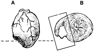
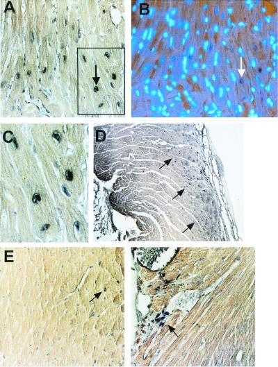
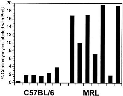
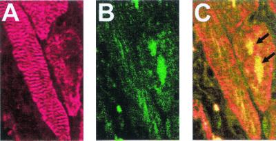
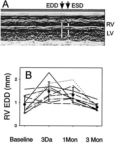
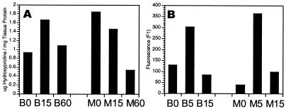
The reaction of cardiac tissue to acute injury involves interacting cascades of cellular and molecular responses that encompass inflammation, hormonal signaling, extracellular matrix remodeling, and compensatory adaptation of myocytes. Myocardial regeneration is observed in amphibians, whereas scar formation characterizes cardiac ventricular wound healing in a variety of mammalian injury models. We have previously shown that the MRL mouse strain has an extraordinary capacity to heal surgical wounds, a complex trait that maps to at least seven genetic loci. Here, we extend these studies to cardiac wounds and demonstrate that a severe transmural, cryogenically induced infarction of the right ventricle heals extensively within 60 days, with the restoration of normal myocardium and function. Scarring is markedly reduced in MRL mice compared with C57BL/6 mice, consistent with both the reduced hydroxyproline levels seen after injury and an elevated cardiomyocyte mitotic index of 10-20% for the MRL compared with 1-3% for the C57BL/6. The myocardial response to injury observed in these mice resembles the regenerative process seen in amphibians.
Figures

Similar articles
-
The MRL mouse repairs both cryogenic and ischemic myocardial infarcts with scar.Cardiovasc Pathol. 2008 Jan-Feb;17(1):14-22. doi: 10.1016/j.carpath.2007.01.007. Epub 2007 Apr 2. Cardiovasc Pathol. 2008. PMID: 18160056
-
Reparative myocardial mechanisms in adult C57BL/6 and MRL mice following injury.Physiol Genomics. 2007 Jun 19;30(1):44-52. doi: 10.1152/physiolgenomics.00070.2006. Epub 2007 Feb 27. Physiol Genomics. 2007. PMID: 17327495
-
Absence of regeneration in the MRL/MpJ mouse heart following infarction or cryoinjury.Cardiovasc Pathol. 2008 Jan-Feb;17(1):6-13. doi: 10.1016/j.carpath.2007.01.005. Epub 2007 Mar 21. Cardiovasc Pathol. 2008. PMID: 18160055 Free PMC article.
-
Spallanzani's mouse: a model of restoration and regeneration.Curr Top Microbiol Immunol. 2004;280:165-89. doi: 10.1007/978-3-642-18846-6_5. Curr Top Microbiol Immunol. 2004. PMID: 14594211 Review.
-
The scarless heart and the MRL mouse.Philos Trans R Soc Lond B Biol Sci. 2004 May 29;359(1445):785-93. doi: 10.1098/rstb.2004.1468. Philos Trans R Soc Lond B Biol Sci. 2004. PMID: 15293806 Free PMC article. Review.
Cited by
-
Brain monoamines and antidepressant-like responses in MRL/MpJ versus C57BL/6J mice.Neuropharmacology. 2013 Apr;67:503-10. doi: 10.1016/j.neuropharm.2012.11.027. Epub 2012 Dec 6. Neuropharmacology. 2013. PMID: 23220293 Free PMC article.
-
Enhanced sensitivity of the MRL/MpJ mouse to the neuroplastic and behavioral effects of chronic antidepressant treatments.Neuropsychopharmacology. 2009 Jun;34(7):1764-73. doi: 10.1038/npp.2008.234. Epub 2009 Jan 28. Neuropsychopharmacology. 2009. PMID: 19177066 Free PMC article.
-
Telomerase expression in the mammalian heart.FASEB J. 2012 Dec;26(12):4832-40. doi: 10.1096/fj.12-208843. Epub 2012 Aug 23. FASEB J. 2012. PMID: 22919071 Free PMC article.
-
Cell-based therapies for the treatment of myocardial infarction: lessons from cardiac regeneration and repair mechanisms in non-human vertebrates.Heart Fail Rev. 2019 Jan;24(1):133-142. doi: 10.1007/s10741-018-9750-8. Heart Fail Rev. 2019. PMID: 30421074 Review.
-
Robust axonal growth and a blunted macrophage response are associated with impaired functional recovery after spinal cord injury in the MRL/MpJ mouse.Neuroscience. 2008 Oct 15;156(3):498-514. doi: 10.1016/j.neuroscience.2008.08.013. Epub 2008 Aug 19. Neuroscience. 2008. PMID: 18786615 Free PMC article.
References
-
- Clark R A F. In: The Molecular and Cellular Biology of Wound Repair. Clark R, editor. New York: Plenum; 1996. pp. 3–35.
-
- Gross J. Wound Repair Regen. 1996;4:190–202. - PubMed
-
- Stocum D L. Wound Repair Regen. 1996;4:3–15. - PubMed
-
- Koishi K, Zhang M, McLennan I S, Harris A J. Dev Dyn. 1995;202:244–254. - PubMed
Publication types
MeSH terms
Substances
Grants and funding
LinkOut - more resources
Full Text Sources
Other Literature Sources
Medical
Molecular Biology Databases
