

Using positron emission tomography with [(18)F]FDG to predict tumor behavior in experimental colorectal cancer
- PMID: 11494112
- PMCID: PMC1505592
- DOI: 10.1038/sj.neo.7900147
Using positron emission tomography with [(18)F]FDG to predict tumor behavior in experimental colorectal cancer
Abstract
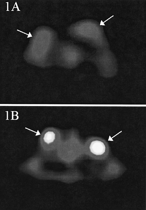
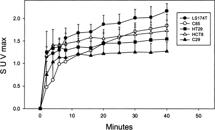
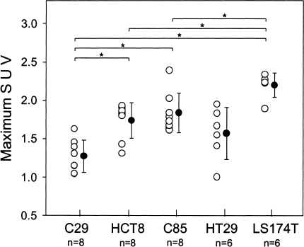
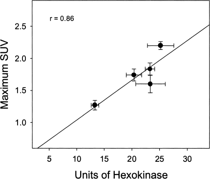
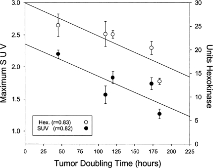
This study investigates the relationship between FDG uptake as determined by positron emission tomography (PET) imaging and rates of tumor growth, cellular GLUT1 transporter density, and the activities of hexokinase and glucose-6-phosphatase in a solid tumor implant model. Five different human colorectal xenografts of different growth properties were implanted in athymic rats and evaluated by dynamic (18)F-FDG-PET. The phosphorylating and dephosphorylating activities of the key glycolytic enzymes, hexokinase and glucose-6-phosphatase, were measured in these tumor types by spectrophotometric assays and the expression of GLUT1 glucose transporter protein was determined by immunohistochemistry. Correlations among FDG accumulation, hexokinase activity, and tumor doubling time are reported in these colon xenografts. The results indicate that the activity of tumor hexokinase may be a marker of tumor growth rate that can be determined by (18)F-FDG-PET imaging. PET scanning may not only be a useful tool for staging patients for extent of disease, but may provide important prognostic information concerning the proliferative rates of malignancies.
Figures
References
-
- Warburg O. On the origin of cancer cells. Science. 1956;123:309–314. - PubMed
-
- Younes M, Lechago LV, Somoano JR, Mosharaf M, Lechago J. Wide expression of the human erythrocyte glucose transporter Glut1 in human cancers. Cancer Res. 1996;56:1164–1167. - PubMed
-
- Brown RS, Leung JY, Fisher SJ, Frey KA, Ethier SP, Wahl RL. Intratumoral distribution of tritiated FDG in breast carcinoma: correlation between Glut-1 expression and FDG uptake. J Nucl Med. 1996;37:1042–1047. - PubMed
-
- Higashi T, Tamaki N, Torizuka T, Nakamoto Y, Sakahara H, Kimura T, Honda T, Inokuma T, Katsushima S, Ohshio G, Imamura M, Konishi J. FDG uptake, GLUT-1 glucose transporter and cellularity in human pancreatic tumors. J Nucl Med. 1998;39:1727–1735. - PubMed
-
- Chung JK, Lee YJ, Kim C, Choi SR, Kim M, Lee K, Jeong JM, Lee DS, Jang JJ, Lee MC. Mechanisms related to [18F] fluorodeoxyglucose uptake of human colon cancers transplanted in nude mice. J Nucl Med. 1999;40:339–346. - PubMed
Publication types
MeSH terms
Substances
Grants and funding
LinkOut - more resources
Full Text Sources
Other Literature Sources
Medical
Miscellaneous