. 2001:1:8.
doi: 10.1186/1472-6793-1-8. Epub 2001 Jul 24.

Modulation of L-type Ca2+ current but not activation of Ca2+ release by the gamma1 subunit of the dihydropyridine receptor of skeletal muscle

Affiliations

Modulation of L-type Ca2+ current but not activation of Ca2+ release by the gamma1 subunit of the dihydropyridine receptor of skeletal muscle

C A Ahern et al. BMC Physiol. 2001.

Abstract

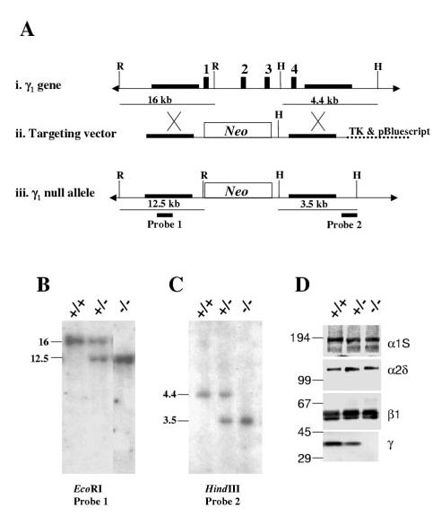
Background: The multisubunit (alpha1S,alpha2-delta, beta1a and gamma1) skeletal muscle dihydropyridine receptor (DHPR) transduces membrane depolarization into release of Ca2+ from the sarcoplasmic reticulum (SR) and also acts as an L-type Ca2+ channel. To more fully investigate the function of the gamma1 subunit in these two processes, we produced mice lacking this subunit by gene targeting.

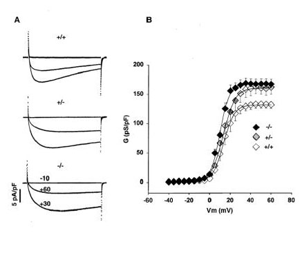
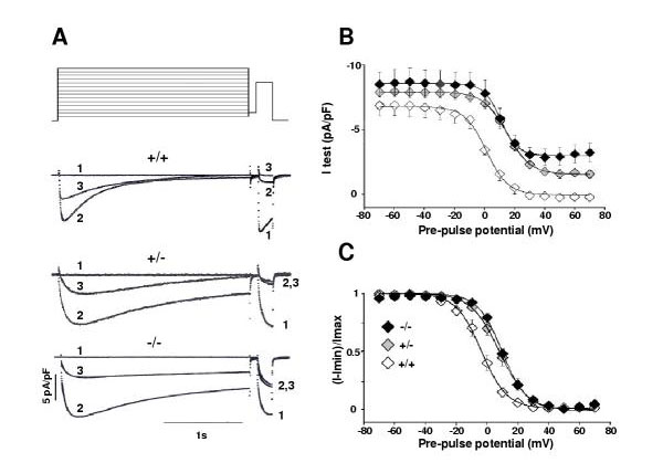
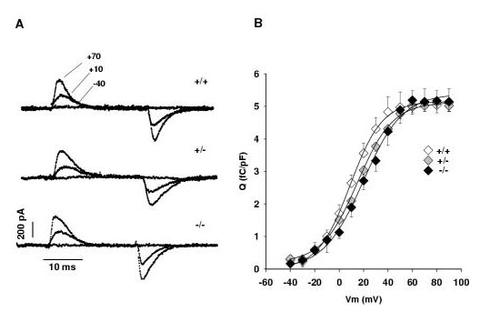
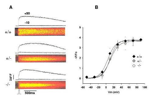
Results: Mice lacking the DHPR gamma1 subunit (gamma1 null) survive to adulthood, are fertile and have no obvious gross phenotypic abnormalities. The gamma1 subunit is expressed at approximately half the normal level in heterozygous mice (gamma1 het). The density of the L-type Ca2+ current in gamma1 null and gamma1 het myotubes was higher than in controls. Inactivation of the Ca2+ current produced by a long depolarization was slower and incomplete in gamma1 null and gamma1 het myotubes, and was shifted to a more positive potential than in controls. However, the half-activation potential of intramembrane charge movements was not shifted, and the maximum density of the total charge was unchanged. Also, no shift was observed in the voltage-dependence of Ca2+ transients. gamma1 null and gamma1 het myotubes had the same peak Ca2+ amplitude vs. voltage relationship as control myotubes.

Conclusions: The L-type Ca2+ channel function, but not the SR Ca2+ release triggering function of the skeletal muscle dihydropyridine receptor, is modulated by the gamma1 subunit.


Figures

Figure 1
Figure 1
Inactivation of the γ1 subunit by gene targeting. A) i. The normal γ1 gene, which contains 4 exons (represented by black boxes). ii. The targeting vector contains a fragment with 4.4 kb of homology to a region 5' of exon 1 and a 2.6 kb fragment with homology to a region 3' of exon 4 (thick horizontal lines). These fragments are separated by the neo gene, which is used to select the ES cells in culture. iii. The modified γ1 locus. When recombination occurs in the regions indicated by the X's, all 4 exons of the γ1 gene are replaced by the neo cassette. B, C) Southern blots of DNA, either digested with EcoRI and hybridized to probe 1 (B), or DNA digested with HindIII and hybridized to probe 2 (C), from control (+/+), γ1 het (+/-) and γ1 null (-/-) mice. These data show the predicted size bands for the wild type and targeted alleles. D) Western blots for each of the four skeletal DHPR subunits from control (+/+), γ1 het (+/-) and γ1 null (-/-) mice. The γ1 subunit is absent from the γ1 null mice and at approximately half the normal level in the γ1 het mice.
Figure 2
Figure 2
L-type Ca2+ conductance in γ1 knockout myotubes. A) Whole-cell L-type Ca2+ current when control (+/+), γ1 het (+/-), or γ1 null (-/-) myotubes are depolarized to either -10 mV, +30 mV or +60 mV from a holding potential of -40 mV. The pulse duration was 500 ms. The cell capacitance was 262 pF, 221 pF, and 286 pF, for the control, γ1 het and γ1 null cells, respectively. B) Voltage dependence of the Ca2+ conductance for 8 control, 8 γ1 het and 12 γ1 null cells. The curves correspond to a Boltzmann fit of the population mean with the following parameters. Gmax = 132 pS/pF, V1/2 = 13.4 mV and k = 5.3 mV for control cells; Gmax = 160 pS/pF, V1/2 = 13 mV and k = 5.8 mV for γ1 het cells; and Gmax = 167 pS/pF, V1/2 = 9.9 mV and k = 4.6 mV for γ1 null cells.
Figure 3
Figure 3
Slow inactivation of the Ca2+ current in γ1 knockout myotubes. A) Diagram to scale of the two-pulse protocol used to inactivate the L-type Ca2+ current at each of 15 potentials and then the test potential (+20 mV) to measure the remaining non-inactivated current. Ca2+ currents during the pre-pulse and test-pulse phases of the protocol are shown for a pre-pulse depolarization to -70 mV (trace 1), +30 mV (trace 2) and +60 mV (trace 3) in a control, a γ1 het, and a γ1 null myotube. The cell capacitance was 317 pF for the control, 175 pF for the γ1 het and 348 pF for the γ1 null cell. B) The maximum Ca2+ current during the test pulse is plotted as a function of the pre-pulse potential for 6 control, 8 γ1 het and 8 γ1 null cells. C) The non-activating component was subtracted and the curves were normalized to show the voltage-dependence of the inactivating component. I is the maximum test current, Imin is the test current at +50 mV and Imax is the test current at -70 mV. The curves correspond to a Boltzmann fit of the population mean with the following parameters. [(I-Imin)/Imax]max = 1, V1/2 = -3.8 mV and k = 8.4 mV for control cells. [(I-Imin)/Imax]max = 1, V1/2 = +15.6 mV and k = 8.1 mV for γ1 het cells. [(I-Imin)/Imax]max = 1, V1/2 = +9.7 mV and k = 7.9 mV for γ1 null cells.
Figure 4
Figure 4
Voltage dependence of intramembrane charge movements in γ1 knockout myotubes. A) Whole-cell current produced by charge movements in response to 3 of 14 25-ms voltage steps delivered to the same cell. The voltage steps shown are to -40 mV, +10 mV, and +70 mV. The cell capacitance was 241 pF for the control, 292 pF for the γ1 het and 253 pF for the γ1 null cell. B) Voltage dependence of charge movement obtained by integration of the OFF transient current for 8 control, 4 γ1 het, and 8 γ1 null cells. The curves correspond to a Boltzmann fit of the population mean with the following parameters. Qmax = 5.2 fC/pF, V1/2 = 13.2 mV and k = 9 mV for control cells. Qmax = 4.9 fC/pF; V1/2 = 15 mV and k = 14.5 mV for γ1 het cells. Qmax = 5.4 fC/pF, V1/2 = 19.6 mV and k = 16 mV for γ1 null cells.
Figure 5
Figure 5
Voltage-dependence of Ca2+ transients in γ1 knockout and normal myotubes. A) Confocal line-scan images of fluo-4 fluorescence in response to a 50 ms step to +90 mV from a holding potential of -40 mV. Hot colors represent high fluorescence (yellow>red). The pulse was delivered 100 ms after the start of the line-scan as indicated at the bottom of the figure. Images have a horizontal dimension of 2.05 seconds in all cases. The vertical dimension was 15, 28, and 24 microns for the control, γ1 het, and γ1 null cells, respectively. The two curves on top of the image show the time course of the fluorescence intensity at -10 mV and +90 mV. 1 ΔF/Fo unit corresponds to a doubling of the cell resting fluorescence. B) Voltage-dependence of the peak ΔF/Fo for 6 control, 4 γ1 het, and 6 γ1 null cells. The curves correspond to a Boltzmann fit of the population mean with the following parameters. ΔF/Fomax = 3.8, V1/2 = 10 mV, k = 9.4 mV for control cells. ΔF/Fomax = 3.6, V1/2 = 10.8 mV; k = 10.9 mV for γ1 het cells. ΔF/Fomax = 3.7, V1/2 = 4.5 mV; k = 10 mV for γ1 null cells.

References

    1. Perez-Reyes E, Schneider T. Calcium channels: Structure, Function, and Classification. Drug Dev Res. 1994;33:295–318.
    1. McPherson PS, Campbell KP. The ryanodine receptor/Ca2+ release channel. J Biol Chem. 1993;268:13765–13768. - PubMed
    1. Rios E, Pizarro G. Voltage sensor of excitation-contraction coupling in skeletal muscle. Physiol Revs. 1991;71:849–908. - PubMed
    1. Tanabe T, Beam KG, Adams BA, Niidome T, Numa S. Regions of the skeletal muscle dihydropyridine receptor critical for excitation-contraction coupling. Nature. 1990;346:567–572. - PubMed
    1. Nakai J, Tanabe T, Konno T, Adams B, Beam KG. Localization in the II-III loop of the dihydropyridine receptor of a sequence critical for excitation-contraction coupling. J Biol Chem. 1998;273:24983–24986. - PubMed

Publication types