. 2001 Oct 5;276(40):37436-42.
doi: 10.1074/jbc.M105725200. Epub 2001 Aug 8.

Proapoptotic stimuli induce nuclear accumulation of glycogen synthase kinase-3 beta

Affiliations

Proapoptotic stimuli induce nuclear accumulation of glycogen synthase kinase-3 beta

G N Bijur et al. J Biol Chem. .

Abstract

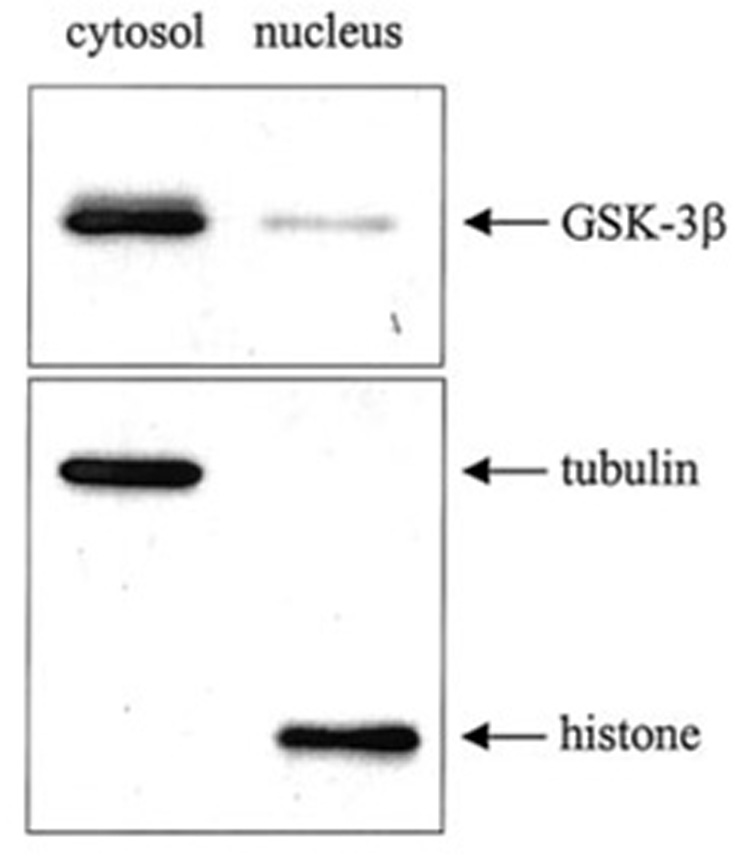
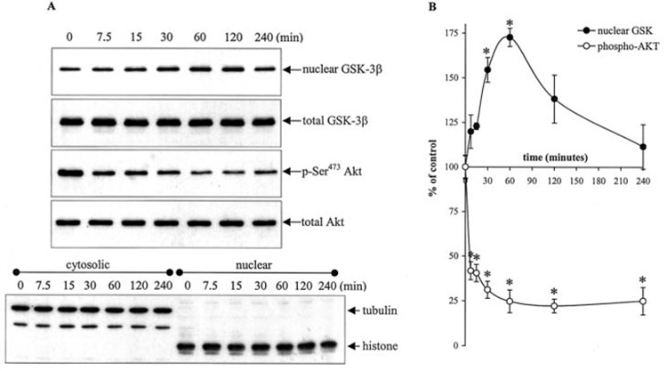
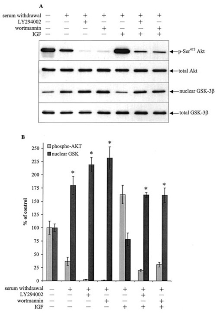
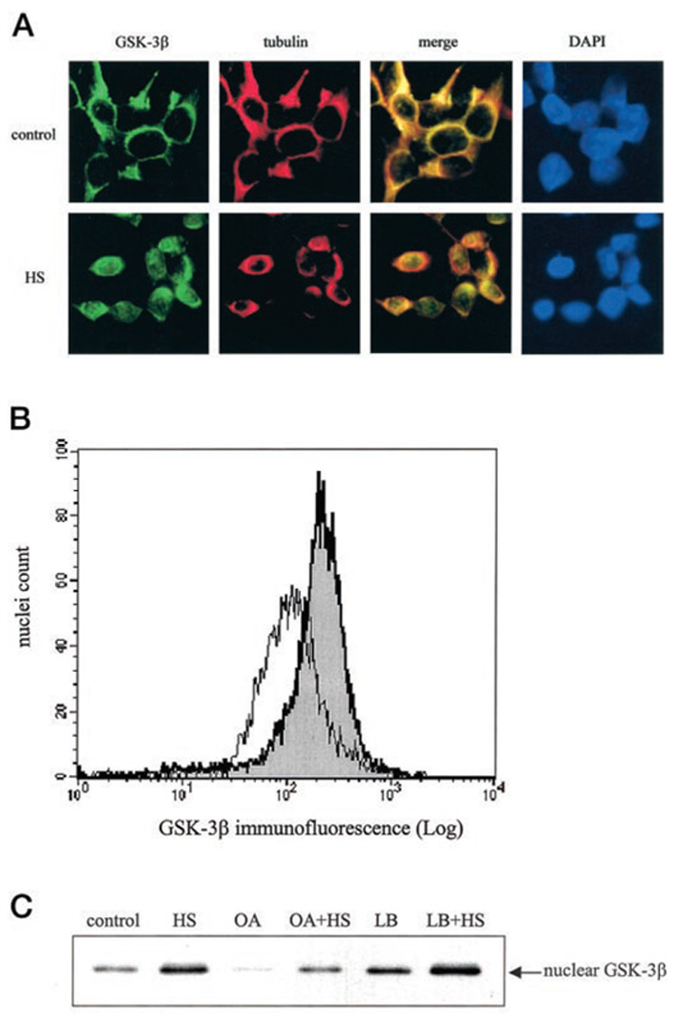
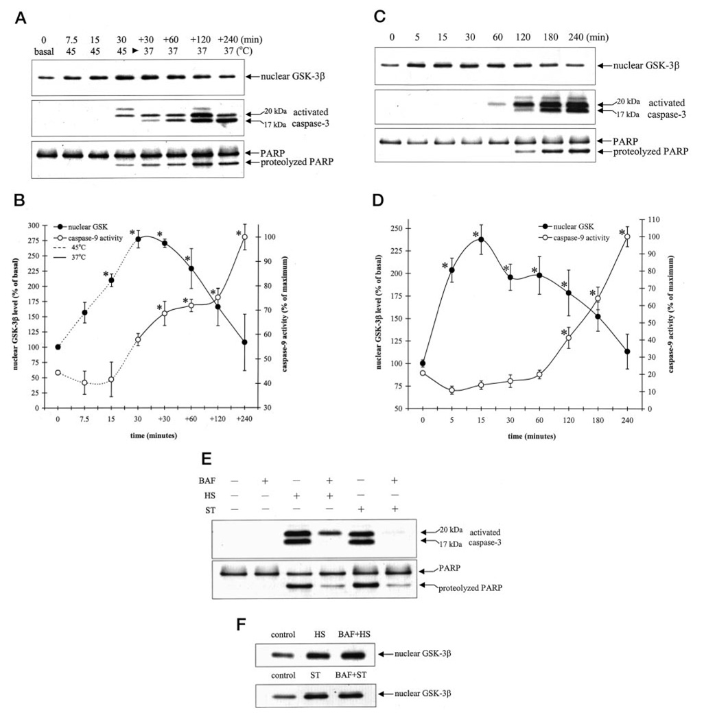
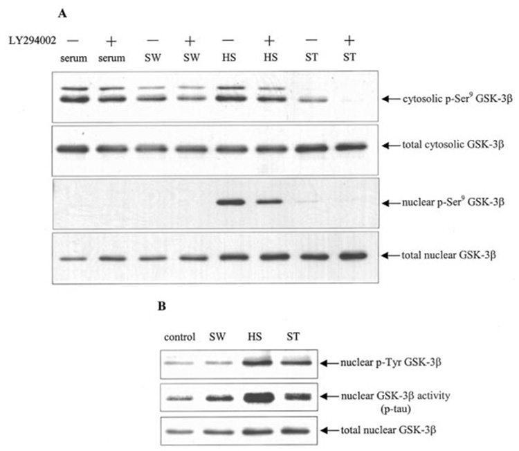
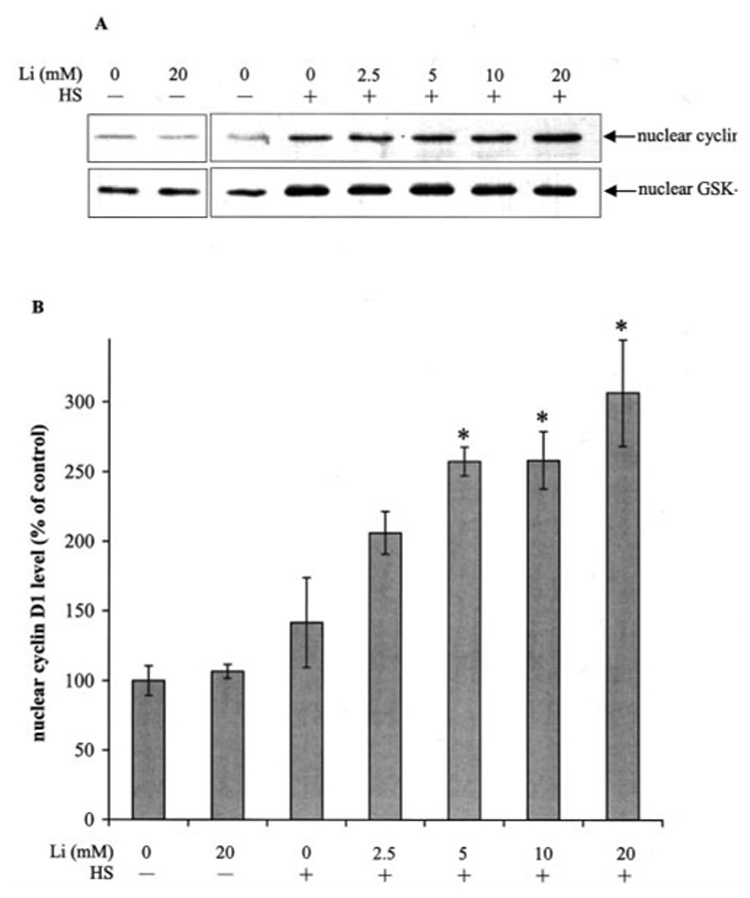
The goal of this study was to determine whether the intracellular distribution of the proapoptotic enzyme glycogen synthase kinase-3 beta (GSK-3 beta) is dynamically regulated by conditions that activate apoptotic signaling cascades. In untreated human neuroblastoma SH-SY5Y cells, GSK-3 beta was predominantly cytosolic, although a low level was also detected in the nucleus. The nuclear level of GSK-3 beta was rapidly increased after exposure of cells to serum-free media, heat shock, or staurosporine. Although each of these conditions caused changes in the serine 9 and/or tyrosine phosphorylation of GSK-3 beta, neither of these modifications was correlated with nuclear accumulation of GSK-3 beta. Heat shock and staurosporine treatments increased nuclear GSK-3 beta prior to activation of caspase-9 and caspase-3, and this nuclear accumulation of GSK-3 beta was unaltered by pretreatment with a general caspase inhibitor. The GSK-3 beta inhibitor lithium did not alter heat shock-induced nuclear accumulation of GSK-3 beta but increased the nuclear level of cyclin D1, indicating that cyclin D1 is a substrate of nuclear GSK-3 beta. Thus, the intracellular distribution of GSK-3 beta is dynamically regulated by signaling cascades, and apoptotic stimuli cause increased nuclear levels of GSK-3 beta, which facilitates interactions with nuclear substrates.


Figures

FIG. 1.
FIG. 1.. GSK-3β levels in the cytoplasm and nucleus
SH-SY5Y cells were fractionated into cytosolic and nuclear fractions as described under “Experimental Procedures.” Extracts (2.5 µg of protein) were immunoblotted for GSK-3β. To verify complete separation of the cytosolic and nuclear fractions, cytosolic and nuclear extracts (10 µg of protein) were immunoblotted for tubulin and histone.
FIG. 2.
FIG. 2.. Serum withdrawal induces nuclear accumulation of GSK-3β
Serum-containing media was withdrawn, and cells were treated with serum-free media for 0, 7.5, 15, 30, 60, 120, and 240 min. One aliquot of cells was fractionated into cytosolic and nuclear fractions, and another aliquot of cells was used to produce total cell lysates as described under “Experimental Procedures.” A, nuclear extracts were immunoblotted for GSK-3β. Cytosolic and nuclear extracts were immunoblotted for tubulin and histone protein to verify complete separation of the cytosolic and nuclear fractions. Total cell lysates were immunoblotted for GSK-3β, p-Ser473 Akt, and total Akt. The blots shown are representative of at least three independent experiments. B, GSK-3β and p-Ser473 Akt bands were analyzed by a scanning densitometer. Values are expressed as a percent of GSK-3β nuclear levels and p-Ser473 Akt levels in untreated cells. Means ± S.E., n = three experiments; *, p < 0.05 (ANOVA) compared with untreated cells.
FIG. 3.
FIG. 3.. PI3K inhibitors and IGF-1 affect serum withdrawal-induced GSK-3β nuclear accumulation
Serum-containing media was withdrawn, and cells were incubated in serum-free media for a total time of 1 h. For combined IGF-1 and LY294002 or wortmannin treatments, cells were incubated in serum-free media for 15 min, treated with 20 µm LY294002 or 40 nm wortmannin for 15 min, and incubated with 50 ng/ml IGF-1 for 30 min. For individual treatments, cells were treated with 20 µm LY294002 or 40 nm wortmannin alone for 45 min or 50 ng/ml IGF-1 alone for 30 min. The nuclear extracts were immunoblotted for GSK-3β. Total cell lysates were immunoblotted for GSK-3β, p-Ser473 Akt, and total Akt. B, nuclear GSK-3β and p-Ser473 Akt bands were analyzed by densitometer. Means ± S.E., n = three experiments; *, p < 0.05 (ANOVA) compared with untreated cells.
FIG. 4.
FIG. 4.. Heat shock induces GSK-3β nuclear accumulation
A, cells cultured on glass coverslips were either maintained in a 37 °C chamber as controls or subjected to heat shock (HS) at 45 °C for 30 min and then transferred to a 37 °C chamber for an additional 30 min. Cells were fixed, permeabilized, and immunofluorescently labeled as described under “Experimental Procedures.” Cells immunofluorescently labeled with GSK-3β-fluorescein isothiocyanate-conjugated antibody fluoresce green, and cells labeled with tubulin-Texas Red-conjugated antibody fluoresce red. Colocalization of GSK-3β and tubulin appears yellow in the merged image. Nuclei were visualized by 4,6-diamino-2-phenylindole (DAPI) staining. A representative section is shown at 400× magnification. B, isolated nuclei from control cells (white peak) and heat shock-treated cells (gray peak) were fixed, immunofluorescently stained with GSK-3β and stained with Hoechst 33342, and analyzed by flow cytometry as described under “Experimental Procedures.” C, control and heat shock-treated (HS) cells were incubated with 100 nM okadaic acid (OA) 1 h prior to heat shock treatment or with 10 ng/ml leptomycin B (LB) 2.5 h prior to heat shock treatment. The nuclear extracts were immunoblotted for GSK-3β.
FIG. 5.
FIG. 5.. Heat shock- and staurosporine-induced GSK-3β nuclear accumulation precedes activation of caspase-9 and caspase-3
Cells were subjected to heat shock at 45 °C for 7.5, 15, and 30 min or subjected to heat shock for 30 min and then transferred to a 37 °C chamber for an additional (+) 30, 60, 120, and 240 min (A and B) or treated with 0.5 µm staurosporine for 5, 15, 30, 60, 120, 180, and 240 min (C and D). Nuclear extracts were immunoblotted for GSK-3β, and total cell lysates were immunoblotted for activated caspase-3 and PARP proteolysis (A and C) or were used to measure caspase-9 activity (B and D) as described under “Experimental Procedures.” Nuclear GSK-3β protein bands were quantitated by densitometer. Means ± S.E., n = three experiments; *, p < 0.05 (ANOVA) compared with untreated cells. E, cells were treated with 100 µm BAF 1 h prior to treatment with 30-min heat shock (HS), followed by incubation at 37 °C for 4 h or with 0.5 µm staurosporine (ST) for 4 h. Total cell lysates were immunoblotted for activated caspase-3 and PARP. F, cells were treated with 100 µm BAF 1 h prior to treatment with 30 min of HS followed by incubation at 37 °C or with 0.5 µm ST for 15 min. Nuclear extracts were immunoblotted for GSK-3β.
FIG. 6.
FIG. 6.. GSK-3β nuclear accumulation is independent of Ser9 and tyrosine phosphorylation
Cells were subjected to 1 h of serum withdrawal (SW), 30 min of heat shock (HS) and incubation at 37 °C for 30 min, or 0.5 µm staurosporine (ST) for 15 min. Where indicated cells were pretreated for 30 min with 20 µm LY294002. A, cytosolic and nuclear extracts were immunoblotted for p-Ser9 GSK-3β and total GSK-3β. B, to measure tyrosine phosphorylation of GSK-3β, nuclear extracts were immunoprecipitated with anti-phosphotyrosine antibody (PY20) and then immunoblotted for GSK-3β to detect tyrosine-phosphorylated (p-Tyr) GSK-3β. For activity measurements, GSK-3β was immunoprecipitated from nuclear extracts, and GSK-3β activity was measured using recombinant tau protein as substrate as described under “Experimental Procedures,” and the immunoprecipitated nuclear GSK-3β was immunoblotted. A representative result from three experiments is shown.
FIG. 7.
FIG. 7.. Lithium treatment enhances heat shock-induced nuclear accumulation of cyclin D1
Cells were incubated with 0 or 20 mm lithium for 90 min or pretreated for 30 min with 0, 2.5, 5, 10, and 20 mm lithium and then treated with 30 min heat shock (HS), followed by incubation in a 37 °C chamber for 30 min. Nuclear extracts were immunoblotted for cyclin D1 and GSK-3β (A), and the cyclin D1 protein bands were quantitated by densitometer (B). Means ± S.E., n = three experiments; *, p < 0.05 (ANOVA) compared with cells treated with heat shock alone.

References

    1. Grimes CA, Jope RS. Prog. Neurobiol. 2001;65:391–426. - PubMed
    1. Wang QM, Fiol CJ, DePaoli-Roach AA, Roach PJ. J. Biol. Chem. 1994;269:14566–14574. - PubMed
    1. Lesort M, Jope RS, Johnson GV. J. Neurochem. 1999;72:576–584. - PubMed
    1. Hartigan JA, Johnson GV. J. Biol. Chem. 1999;274:21395–21401. - PubMed
    1. Alessi DR, James SR, Downes CP, Holmes AB, Gaffney PR, Reese CB, Cohen P. Curr. Biol. 1997;7:261–269. - PubMed

Publication types