

Decline of CD3-positive T-cell counts by 6 months of age is associated with rapid disease progression in HIV-1--infected infants
- PMID: 11496244
- PMCID: PMC4357165
- DOI: 10.1067/mai.2001.116573
Decline of CD3-positive T-cell counts by 6 months of age is associated with rapid disease progression in HIV-1--infected infants
Abstract
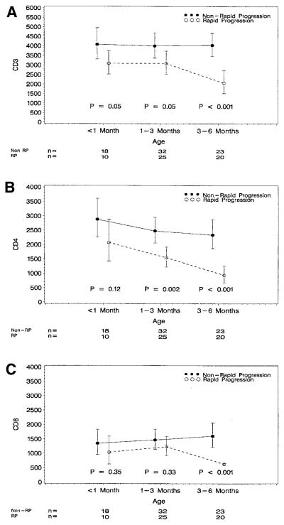
Because HIV-1 infected infants with rapid progression (RP) of disease might benefit from early and intense antiretroviral therapy, the identification of predictive factors of RP becomes extremely important. Currently, the best predictive factors of RP in HIV-1 infected children are HIV-1 RNA levels and CD4-positive T-cell counts. A decrease in CD3-positive T-cell count has been identified as a predictive factor of AIDS development in HIV-1 infected adults. Our objective was to evaluate decreased number of CD3-positive T-cells as a predictive factor of RP in infants. Peripheral blood lymphocytes from HIV-1 infected infants (up to 6 months of age) were analyzed for an association of lymphocyte subsets with RP, which was defined as the occurrence of AIDS or death before 18 months of age. In infants with RP (n = 32), CD3-positive T-cell counts were 3093 cells/microL at <1 month of age, 3092 cells/microL at 1 to 3 months, and 2062 cells/microL at 3 to 6 months. Non-RP infants (n = 49) maintained their CD3-positive T-cells counts at approximately 4000 cells/microL for at least 6 months of life. CD3-positive and CD4-positive T-cell counts were significantly associated with RP. Our results suggest that a decreased CD3-positive T-cell count may be used to predict RP in HIV-1 infected infants (RR = 2.16, P =.001).
Figures
References
-
- Centers for Disease Control. US HIV-1-AIDS cases reported through June 1999. Atlanta: CDC; HIV-1/AIDS surveillance report 1999; pp. 1–28.
-
- Centers for Disease Control and Prevention. 1994 revised classification for human immunodeficiency virus infection in children less than 13 years of age. Mor Mortal Wkly Rep CDC Surveill Summ. 1994;43:1–10.
-
- Mayaux M-J, Burgard M, Teglas J-P, et al. Neonatal characteristics in rapidly progressive perinatally acquired HIV-1 disease. JAMA. 1996;275:606–10. - PubMed
-
- MMWR. Guidelines for use of antiretroviral agents in pediatrics HIV-1 infection. Mor Mortal Wkly Rep. 1998;47:1–43.
-
- Mofenson LM, Korelitz J, Meyer WA, et al. The relationship between serum human immunodeficiency virus type 1 (HIV-1-1) RNA level, CD4-positive lymphocyte percent, and long term mortality risk in HIV-1-1 infected children. J Infect Dis. 1997;175:1029–38. - PubMed
Publication types
MeSH terms
Substances
Grants and funding
- K01 RR000188/RR/NCRR NIH HHS/United States
- M01 RR000645/RR/NCRR NIH HHS/United States
- N01-HR-96039/HR/NHLBI NIH HHS/United States
- N01-HR-96040/HR/NHLBI NIH HHS/United States
- RR-00071/RR/NCRR NIH HHS/United States
- RR-00643/RR/NCRR NIH HHS/United States
- M01 RR000865/RR/NCRR NIH HHS/United States
- M01 RR000533/RR/NCRR NIH HHS/United States
- N01 HR096037/HR/NHLBI NIH HHS/United States
- M01 RR002172/RR/NCRR NIH HHS/United States
- M01 RR000188/RR/NCRR NIH HHS/United States
- M01 RR000071/RR/NCRR NIH HHS/United States
- RR-00188/RR/NCRR NIH HHS/United States
- RR-02172/RR/NCRR NIH HHS/United States
- RR-00533/RR/NCRR NIH HHS/United States
- RR-00645/RR/NCRR NIH HHS/United States
- RR-00865/RR/NCRR NIH HHS/United States
- N01-HR-96042/HR/NHLBI NIH HHS/United States
- N01-HR-96038/HR/NHLBI NIH HHS/United States
- N01-HR-96041/HR/NHLBI NIH HHS/United States
- N01 HR096043/HR/NHLBI NIH HHS/United States
LinkOut - more resources
Full Text Sources
Medical
Research Materials
