

Detection of intracranial hemorrhage: comparison between gradient-echo images and b(0) images obtained from diffusion-weighted echo-planar sequences
- PMID: 11498414
- PMCID: PMC7975215
Detection of intracranial hemorrhage: comparison between gradient-echo images and b(0) images obtained from diffusion-weighted echo-planar sequences
Abstract
Background and purpose: Diffusion-weighted MR imaging (DWI) is commonly used as the initial and sole imaging examination for the detection of acute cerebral infarction, yet it remains controversial whether MR can detect hyperacute (<24 h) hemorrhage. Hemorrhage is best detected with gradient-echo (GRE) T2*-weighted sequences, because of their magnetic susceptibility effects. DWI uses a spin-echo echo-planar technique (EPI) that is more sensitive than spin-echo T2-weighted imaging to susceptibility effects. Our aim was to determine whether the b(0) image from the DWI-EPI sequence is as sensitive as GRE in detecting hemorrhagic lesions on imaging studies performed to identify acute infarction or hemorrhage.
Methods: All MR studies performed for clinically suspected or radiographically confirmed acute infarction or hemorrhage from 2/1/98 to 8/15/99 were retrospectively interpreted by one neuroradiologist in a blinded fashion. The sensitivity of hemorrhage detection, conspicuity of lesions, and diagnostic certainty were compared between the b(0) EPI and GRE sequences.
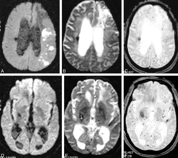
Results: We found 101 acute infarcts, of which 13 were hemorrhagic, as evidenced by the presence of hypointensity within the infarction on the GRE sequence. This finding served as the reference standard for detection of hemorrhage. Hemorrhage was diagnosed with confidence in only seven cases (54%) on b(0) images; 22 acute hematomas were hypointense on GRE images whereas 19 were hypointense on b(0) images (86%); 17 chronic hematomas were depicted on GRE images and 12 on b(0) scans (63%). Punctate hemorrhages and linear cortical staining were detected on 37 GRE studies but on only four b(0) studies. Hemorrhage was always more conspicuous on the GRE sequences.
Conclusion: b(0) images from a DWI sequence failed to detect minimally hemorrhagic infarctions and small chronic hemorrhages associated with microangiopathy. GRE scans were more sensitive than b(0) images in the detection of these hemorrhages and should be included in emergency brain MR studies for acute infarction, especially when thrombolytic therapy is contemplated.
Figures


References
-
- Moseley ME, Cohen Y, Mintorovitch J, et al. Early detection of regional cerebral ischemia in cats: comparison of diffusion- and T2-weighted MRI and spectroscopy. Magn Reson Med 1990;14:330-346 - PubMed
-
- Gonzalez RG, Schaefer PW, Buonanno FS, et al. Diffusion-weighted MR imaging: diagnostic accuracy in patients imaged within 6 hours of stroke symptom onset. Radiology 1999;210:155-162 - PubMed
-
- Uluğ AM, Beauchamp N, Bryan RN, van Zijl PCM. Absolute quantitation of diffusion constants in human stroke. Stroke 1997;28:483-490 - PubMed
-
- Beauchamp N, Bryan RN. Acute cerebral ischemic infarction: a pathophysiologic review and radiologic perspective. AJR Am J Roentgenol 1998;171:73-84 - PubMed
-
- Zimmerman RD, Heier LA, Snow RB, Liu DP, Kelly AB, Deck MDF. MR imaging feature of acute intracranial hemorrhage studied at 0.5 T with emphasis on sequential intensity changes on multiple pulse sequences. AJNR Am J Neuroradiol 1988;9:47-57
Publication types
MeSH terms
LinkOut - more resources
Full Text Sources