. 2001 Aug;22(7):1316-24.

Proton MR spectroscopic evaluation of suspicious brain lesions after stereotactic radiotherapy

Affiliations

Proton MR spectroscopic evaluation of suspicious brain lesions after stereotactic radiotherapy

H P Schlemmer et al. AJNR Am J Neuroradiol. 2001 Aug.

Abstract

Background and purpose: The radiologic assessment of suspicious brain lesions after stereotactic radiotherapy of brain tumors is difficult. The purpose of our study was to define parameters from single-voxel proton MR spectroscopy that provide a probability measure for differentiating neoplastic from radiation-induced, nonneoplastic lesions.

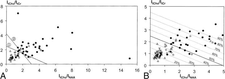
Methods: Seventy-two lesions in 56 patients were examined using a combined MR imaging and MR spectroscopy protocol (point-resolved spectroscopy, TE = 135 ms). Signal intensities of cholines, creatines, N-acetyl aspartate, and the presence of lactate and lipid resonances were correlated to final diagnoses established by clinical and MR imaging follow-up, positron emission tomography studies, or biopsy/surgery. Statistical analysis was performed using the t test, linear discriminant analysis, and k nearest-neighbor method.

Results: Significantly increased signal intensity ratios I(tCho)/I(tCr) (P <.0001) and I(tCho)/I(NAA) (P <.0001) were observed in neoplastic (n = 34) compared with nonneoplastic lesions (n = 32) and contralateral normal brain (n = 33). Analysis of I(tCho)/I(tCr) and I(tCho)/I(NAA) data yielded correct retrospective classification as neoplastic and nonneoplastic in 82% and 81% of the lesions, respectively. Neither I(NAA)/I(tCr) nor signal intensitities of lactate or lipids were useful for differential diagnosis.

Conclusion: Metabolic information provided by proton MR spectroscopy is useful for the differentiation of neoplastic and nonneoplastic brain lesions after stereotactic radiotherapy of brain tumors.


Figures

<sc>fig</sc> 1.
fig 1.
A 67-year-old patient with astrocytoma grade II. MR examination 36 months after stereotactic radiotherapy showed two contrast-enhancing lesions on T1-weighted spin-echo images. A and C, A lesion with ring-shaped enhancement zone is seen in the left frontal lobe (A) and a lesion with homogeneous enhancement in seen in the left temporal lobe (C) (boxes indicate MRS voxels). B and D, 1H MR spectra [double spin-echo sequence 1500/135/200 (TR/TE/excitations)] of both lesions show a peak attributed to free lipids, indicating necrosis. An intense tCho resonance is only seen in the lesion in the left temporal lobe. FDG-PET revealed high glucose uptake in this region, indicating tumor progression. Low tCho signal was found in the lesion in the left frontal lobe. Radionecrosis in this region was confirmed by biopsy.
<sc>fig</sc> 2.
fig 2.
A 36-year-old patient with astrocytoma II in the right temporo-parietal lobe, examined before (A) and 20 months after stereotactic radiotherapy (B). During follow-up, an irregular contrast-enhancing lesion appeared at the upper region of the irradiated volume (B) with complete involution 13 months later (D). 1H MR spectra (double spin-echo sequence 1500/135/200) (C) show initially elevated ItCho/ItCr and ItCho/INAA values and a strong lipid resonance. Thirteen months later, the spectrum indicates normal brain tissue (E)
<sc>fig</sc> 3.
fig 3.
A 54-year-old patient with astrocytoma II of the right temporal lobe after surgery and stereotactic radiotherapy. T2-weighted turbo spin-echo images obtained before (A) and 9 months after radiotherapy (B) show a hyperintense lesion within the irradiated insula on the right hemisphere, a surgical tissue defect in the right temporal, and gliosis in the right occipital lobe (T1-weighted spin-echo images showed no uptake of contrast medium.) Corresponding 1H MR spectra (double spin-echo sequence 1500/135/200) acquired 9, 15, and 21 months after stereotactic radiotherapy (C–E) demonstrated similar signal intensities of tCho and tCr, with only minor changes during follow-up. MR imaging follow-up showed no further change of the lesion consistent with diagnosis of nonneoplastic lesion
<sc>fig</sc> 4.
fig 4.
a, Ratios of 1H MR signal intensities (double spin-echo sequence 1500/135/200–300) of total choline and total creatine (ItCho/ItCr) versus ratios of intensities of total choline and NAA (ItCho/INAA) observed in lesions classified as progressive tumor (●, n = 35), radiation injury (Δ, n = 17), and stable disease (⋄, n = 15). The straight lines: (ItCho/ItCr) = 2.5–0.5 × (ItCho/INAA) (line 1) and (ItCho/ItCr) = 4.2–1.35 × (ItCho/INAA) (line 2) obtained from linear discriminant analysis and 7-point neighbor method, respectively, differentiate neoplastic and nonneoplastic lesions. b, Detail of (a) with ratios ≤ 5.0 together with ItCho/ItCr and ItCho/INAA values of normal brain (×, n = 33, voxel in the contralateral unaffected side). Parallel lines obtained from linear discriminant analysis are labeled with the probability of PT.

Similar articles

Cited by

References

    1. van Kampen M, Engenhart-Cabillic R, Debus J, Hess T, Schad LR, Wannenmacher MF. Low-grade astrocytoma: treatment with conventionally fractionated stereotactic radiation therapy. Radiology 1996;201:275-278 - PubMed
    1. Schultheiss TE, Kun LE, Ang KK, Stephens LC. Radiation response of the central nervous system. Int J Radiat Oncol Biol Phys 1995;31:1093-1112 - PubMed
    1. Sheline GE, Wara WM, Smith V. Therapeutic irradiation and brain injury. Int J Radiat Oncol Biol Phys 1980;6:1215-1228 - PubMed
    1. Frahm J, Bruhn H, Gyngell ML, Merboldt KD, Hänicke W, Sauter R. Localized high–resolution proton NMR spectroscopy using stimulated echoes: initial applications to human brain in vivo. Magn Reson Med 1989;9:79-93 - PubMed
    1. Ross B, Michaelis T. Clinical applications of magnetic resonance spectroscopy. Magn Reson Q 1994;10:191-247 - PubMed

Publication types

MeSH terms