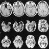
Detection of mesial temporal lobe hypoperfusion in patients with temporal lobe epilepsy by use of arterial spin labeled perfusion MR imaging
- PMID: 11498422
- PMCID: PMC7975208
Detection of mesial temporal lobe hypoperfusion in patients with temporal lobe epilepsy by use of arterial spin labeled perfusion MR imaging
Abstract
Background and purpose: Interictal hypometabolism has lateralizing value in cases of temporal lobe epilepsy and positive predictive value for seizure-free outcome after surgery to treat epilepsy. Alterations in regional cerebral metabolism can also be inferred from measurements of regional cerebral perfusion. The purpose of this study was to determine the feasibility of detecting cerebral blood flow (CBF) asymmetries in the mesial temporal lobes using continuous arterial spin labeling perfusion MR imaging, which is a noninvasive method for calculating regional CBF.
Methods: Twelve patients with medically refractory temporal lobe epilepsy who underwent preoperative evaluation for temporal lobectomy and 12 normal control participants were studied retrospectively. Absolute and normalized mesial temporal CBF measurements were compared between the patient and control groups. Lateralization based on a perfusion asymmetry index was compared with metabolic ((18)[F]-fluorodeoxyglucose positron emission tomography) and hippocampal volumetric asymmetry indices and with clinical lateralization.
Results: Mesial temporal CBF was more asymmetric in patients with temporal lobe epilepsy than in normal control participants, although asymmetric mesial temporal CBF was also found in normal participants, with the left side dominant. Ipsilateral mesial temporal CBF was significantly decreased compared with contralateral mesial temporal CBF in patients with temporal lobe epilepsy. Global CBF measurements were significantly decreased in patients compared with control participants. Asymmetry in mesial temporal blood flow in patients persisted after normalization to global CBF. Lateralization using continuous arterial spin labeling perfusion MR imaging asymmetry index significantly correlated with lateralization based on (18)[F]-fluorodeoxyglucose positron emission tomography hypometabolism, hippocampal volumes, and clinical evaluation.
Conclusion: Continuous arterial spin labeling perfusion MR imaging can detect interictal asymmetries in mesial temporal lobe perfusion in patients with temporal lobe epilepsy. This technique is readily combined with routine structural assessment and potentially offers an inexpensive and noninvasive means of screening for asymmetries in interictal mesial temporal lobe function.
Figures

Similar articles
-
Usefulness of pulsed arterial spin labeling MR imaging in mesial temporal lobe epilepsy.Epilepsy Res. 2008 Dec;82(2-3):183-9. doi: 10.1016/j.eplepsyres.2008.08.001. Epilepsy Res. 2008. PMID: 19041041 Free PMC article. Clinical Trial.
-
Neocortical temporal FDG-PET hypometabolism correlates with temporal lobe atrophy in hippocampal sclerosis associated with microscopic cortical dysplasia.Epilepsia. 2003 Apr;44(4):559-64. doi: 10.1046/j.1528-1157.2003.36202.x. Epilepsia. 2003. PMID: 12681005
-
Correlations of interictal FDG-PET metabolism and ictal SPECT perfusion changes in human temporal lobe epilepsy with hippocampal sclerosis.Neuroimage. 2006 Aug 15;32(2):684-95. doi: 10.1016/j.neuroimage.2006.04.185. Epub 2006 Jun 9. Neuroimage. 2006. PMID: 16762567
-
Mesial temporal lobe epilepsy - An overview of surgical techniques.Int J Surg. 2016 Dec;36(Pt B):411-419. doi: 10.1016/j.ijsu.2016.10.027. Epub 2016 Oct 20. Int J Surg. 2016. PMID: 27773861 Review.
-
Arterial spin-labeling in routine clinical practice, part 2: hypoperfusion patterns.AJNR Am J Neuroradiol. 2008 Aug;29(7):1235-41. doi: 10.3174/ajnr.A1033. Epub 2008 Mar 20. AJNR Am J Neuroradiol. 2008. PMID: 18356467 Free PMC article. Review.
Cited by
-
Shared cognitive and behavioral impairments in epilepsy and Alzheimer's disease and potential underlying mechanisms.Epilepsy Behav. 2013 Mar;26(3):343-51. doi: 10.1016/j.yebeh.2012.11.040. Epub 2013 Jan 13. Epilepsy Behav. 2013. PMID: 23321057 Free PMC article. Review.
-
Resting-state fMRI studies in epilepsy.Neurosci Bull. 2012 Aug;28(4):449-55. doi: 10.1007/s12264-012-1255-1. Neurosci Bull. 2012. PMID: 22833042 Free PMC article. Review.
-
Role of Interictal Arterial Spin Labeling Magnetic Resonance Perfusion in Mesial Temporal Lobe Epilepsy.Ann Indian Acad Neurol. 2021 Jul-Aug;24(4):495-500. doi: 10.4103/aian.AIAN_1274_20. Epub 2021 May 28. Ann Indian Acad Neurol. 2021. PMID: 34728940 Free PMC article.
-
MRI perfusion analysis using freeware, standard imaging software.BMC Vet Res. 2020 May 18;16(1):141. doi: 10.1186/s12917-020-02352-0. BMC Vet Res. 2020. PMID: 32423403 Free PMC article.
-
Application of Arterial Spin Labelling in the Assessment of Ocular Tissues.Biomed Res Int. 2016;2016:6240504. doi: 10.1155/2016/6240504. Epub 2016 Mar 15. Biomed Res Int. 2016. PMID: 27066501 Free PMC article. Review.
References
-
- Duncan J. Imaging and epilepsy. Brain 1997;120:339-377 - PubMed
-
- Henry T, Engel J Jr, Mazziotta J. Clinical evaluation of interictal fluorine-18-fluorodeoxyglucose PET in partial epilepsy. J Nucl Med 1993;34:1892-1898 - PubMed
-
- Weinand M, Carter L. Surface cortical cerebral blood flow monitoring and single photon emission computed tomography: prognostic factors for selecting temporal lobectomy candidates. Seizure 1994;3:55-59 - PubMed
-
- Manno E, Sperling M, Ding X, et al. Predictors of outcome after anterior temporal lobectomy: positron emission tomography. Neurology 1994;44:2331-2336 - PubMed
Publication types
MeSH terms
Substances
Grants and funding
LinkOut - more resources
Full Text Sources
Medical
