

Case Reports
Intraosseous hemangioma of the zygoma: CT and MR findings
Affiliations
- PMID: 11498432
- PMCID: PMC7975228
Item in Clipboard
Case Reports
Intraosseous hemangioma of the zygoma: CT and MR findings
AJNR Am J Neuroradiol.
2001 Aug.
Abstract
Intraosseous hemangiomas are uncommon, constituting less than 1% of all osseous tumors. The most frequent sites are the calvaria and the vertebral column. Involvement of the facial bones is rare, and occurs most commonly in the maxilla, mandible, and nasal bones. Only 20 cases of zygomatic involvement have been reported in the English-language literature. We report a case of an intraosseous hemangioma of the zygoma documented by CT and MR studies.
Figures
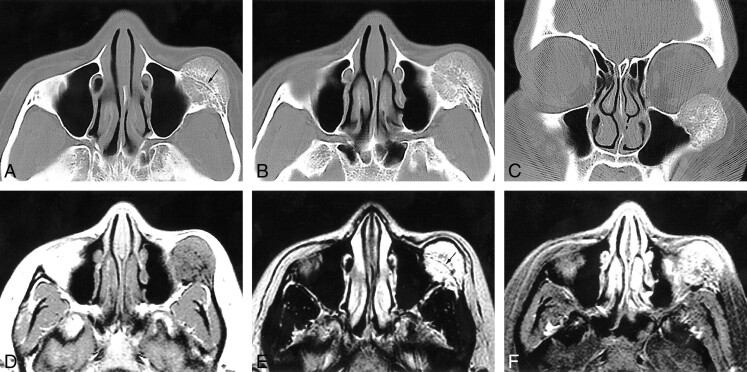
31-year-old woman with a 3-month history of progressive dystopia and a 1-year history of painless swelling of the left cheek. A–C, Axial caudal (A), cranial (B), and coronal (C) CT scans, viewed at wide window settings. A, A 2.5-cm round lesion arises from the zygoma. The inner and outer cortices, although thinned, are preserved. The trabeculae radiate in a spokewheel-type pattern, and a residual trace of the original cortex is seen (arrow). B, The trabeculae of the lesion taper into the zygomatic arch. There is no associated soft tissue mass. C, The rounded lesion encroaches on the inferolateral orbit. D–F, Axial T1-weighted (500/9/2) (D), T2-weighted (3600/95/2) (E), and contrast-enhanced, fat-suppressed T1-weighted (450/9/2) (F) MR images. D, The lesion is isointense with muscle and shows fine radiating lines of high signal intensity that are interspersed with the trabecular signal voids. E, Encroachment on the inferolateral aspect of the orbit is seen. No extraosseous mass is evident. A line of signal representing the original cortex is visible (arrow). F, The mass enhances; no large regional vessels are evident.
References
-
- Clauser L, Meneghini A, Riga M, et al. Haemangioma of the zygoma: report of two cases with a review of the literature. J Craniomaxillofac Surg 1991;19:353-358 - PubMed
-
- Cuesta-Gil M, Navarro-Villa V. Intraosseous hemangioma of the zygomatic bone: a case report. Int J Oral Maxillofac Surg 1992;21:287-291 - PubMed
-
- Har-El G, Hadar T, Zirkin HY, et al. Hemangioma of the zygoma. Ann Plast Surg 1987;18:533-540 - PubMed
-
- Jeter TS, Hackney FL, Aufdemorte TB. Cavernous hemangioma of the zygoma: report of cases. J Oral Maxillofac Surg 1990;48:508-512 - PubMed
-
- Pinna V, Clauser L, Marchi M, Castellan L. Haemangioma of the zygoma: a case report. Neuroradiology 1997;39:216-218 - PubMed
Publication types
MeSH terms
LinkOut - more resources
Full Text Sources
Medical