Unusual CT and MR findings of inflammatory pseudotumor in the parapharyngeal space: case report
- PMID: 11498435
- PMCID: PMC7975212
Unusual CT and MR findings of inflammatory pseudotumor in the parapharyngeal space: case report
Abstract
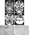
Unusual MR and CT findings of an inflammatory pseudotumor in the parapharyngeal space of a 73-year-old woman are reported. The mass was hypointense on T1- and T2-weighted images and demonstrated ring enhancement after contrast medium injection. Punctated calcifications were scattered at the periphery. Inflammatory pseudotumors in the parapharyngeal space are rare, and only three cases have been reported. The possible pathogenesis and varieties of inflammatory pseudotumors are discussed.
Figures

Similar articles
-
Inflammatory pseudotumor of the spleen: CT and MRI findings.Int Surg. 2007 Mar-Apr;92(2):119-22. Int Surg. 2007. PMID: 17518256
-
Castleman's disease in the retropharyngeal space: CT and MR imaging findings.AJNR Am J Neuroradiol. 2000 Aug;21(7):1337-9. AJNR Am J Neuroradiol. 2000. PMID: 10954291 Free PMC article.
-
Inflammatory pseudotumor of the parapharyngeal space: a case report.Auris Nasus Larynx. 2010 Jun;37(3):397-400. doi: 10.1016/j.anl.2009.08.002. Epub 2009 Oct 25. Auris Nasus Larynx. 2010. PMID: 19857937
-
Inflammatory pseudotumor of the parapharyngeal space: case report and review of the literature.Head Neck. 1992 May-Jun;14(3):230-4. doi: 10.1002/hed.2880140311. Head Neck. 1992. PMID: 1587741 Review.
-
Inflammatory pseudotumor in the epidural space of the thoracic spine: a case report and literature review of MR imaging findings.AJNR Am J Neuroradiol. 2005 Nov-Dec;26(10):2667-70. AJNR Am J Neuroradiol. 2005. PMID: 16286421 Free PMC article. Review.
Cited by
-
Parapharyngeal Angiofibroma: A Case Report.Iran J Radiol. 2015 Jul 22;12(3):e17353. doi: 10.5812/iranjradiol.12(3)2015.17353. eCollection 2015 Jul. Iran J Radiol. 2015. PMID: 26557274 Free PMC article.
-
Inflammatory myofibroblastic tumor of the orbit with associated enhancement of the meninges and multiple cranial nerves.AJNR Am J Neuroradiol. 2006 Nov-Dec;27(10):2217-20. AJNR Am J Neuroradiol. 2006. PMID: 17110698 Free PMC article.
-
A rare case of inflammatory pseudotumour of the submandibular lymphnode.Indian J Otolaryngol Head Neck Surg. 2006 Oct;58(4):408-9. doi: 10.1007/BF03049616. Indian J Otolaryngol Head Neck Surg. 2006. PMID: 23120369 Free PMC article.
-
Prognostic factors in adult brainstem glioma: a tertiary care center analysis and review of the literature.J Neurol. 2022 Mar;269(3):1574-1590. doi: 10.1007/s00415-021-10725-0. Epub 2021 Aug 3. J Neurol. 2022. PMID: 34342680 Free PMC article. Review.
-
Diagnosis and Treatment of Inflammatory Pseudotumor with Lower Cranial Nerve Neuropathy by Endoscopic Endonasal Approach: A Systematic Review.Diagnostics (Basel). 2022 Sep 3;12(9):2145. doi: 10.3390/diagnostics12092145. Diagnostics (Basel). 2022. PMID: 36140546 Free PMC article. Review.
References
-
- Hytiroglou P, Brandwein MS, Strauchen JA, Mirante JP, Urken ML, Biller HF. Inflammatory pseudotumor of the parapharyngeal space: case report and review of the literature. Head Neck 1992;14:230-234 - PubMed
-
- Chan Y-F, Tung M, Yeung CK, Lam KH. Parapharyngeal inflammatory pseudotumor presenting as fever of unknown origin in a 3-year-old girl. Pediatr Pathol 1988;8:195-203 - PubMed
-
- Wold LE, Weiland LH. Tumefactive fibro-inflammatory lesions of the head and neck. Am J Surg Pathol 1983;7:477-482 - PubMed
-
- Coffin CM, Watterson J, Priest JR, Dehner LP. Extrapulmonary inflammatory myofibroblastic tumor (inflammatory pseudotumor): a clinicopathologic and immunohistochemical study of 84 cases. Am J Surg Pathol 1995;19:859-872 - PubMed
Publication types
MeSH terms
LinkOut - more resources
Full Text Sources
Medical
