. 2001 Aug 15;20(16):4370-9.
doi: 10.1093/emboj/20.16.4370.

Regulation of elongation factor 2 kinase by p90(RSK1) and p70 S6 kinase

Affiliations

Regulation of elongation factor 2 kinase by p90(RSK1) and p70 S6 kinase

X Wang et al. EMBO J. .

Abstract

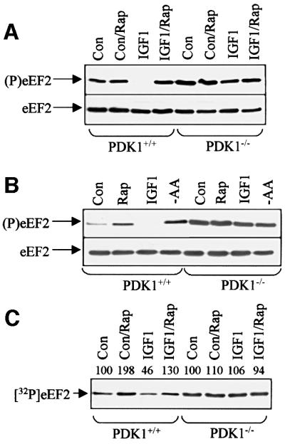
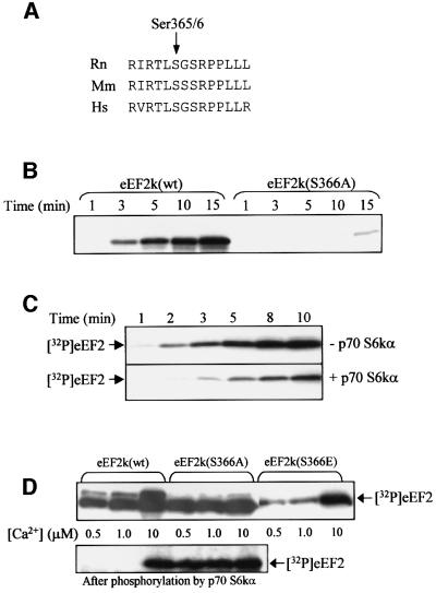
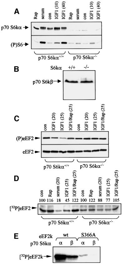
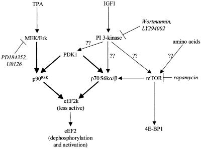
Elongation factor 2 kinase (eEF2k) phosphorylates and inactivates eEF2. Insulin induces dephosphorylation of eEF2 and inactivation of eEF2 kinase, and these effects are blocked by rapamycin, which inhibits the mammalian target of rapamycin, mTOR. However, the signalling mechanisms underlying these effects are unknown. Regulation of eEF2 phosphorylation and eEF2k activity is lost in cells in which phosphoinositide-dependent kinase 1 (PDK1) has been genetically knocked out. This is not due to loss of mTOR function since phosphorylation of another target of mTOR, initiation factor 4E-binding protein 1, is not defective. PDK1 is required for activation of members of the AGC kinase family; we show that two such kinases, p70 S6 kinase (regulated via mTOR) and p90(RSK1) (activated by Erk), phosphorylate eEF2k at a conserved serine and inhibit its activity. In response to insulin-like growth factor 1, which activates p70 S6 kinase but not Erk, regulation of eEF2 is blocked by rapamycin. In contrast, regulation of eEF2 by stimuli that activate Erk is insensitive to rapamycin, but blocked by inhibitors of MEK/Erk signalling, consistent with the involvement of p90(RSK1).


Figures

None
Fig. 1. Regulation of eEF2 and eEF2k in ES cells. (A) ES cells (PDK1+/+ or PDK1–/– as indicated, >80% confluent) were serum starved for 3 h and then treated with rapamycin (Rap) and/or IGF1 (40 min) as shown. Samples were subjected to SDS–PAGE and western blotting. Upper section: antibody against eEF2 phosphorylated at Thr56; lower section: blot with antibody detecting eEF2 irrespective of its phosphorylation state (loading control). (B) PDK+/+ or PDK1–/– ES cells (∼70% confluent) were serum starved for 3 h and then treated with rapamycin (Rap), IGF1 (20 min) or transferred to D-PBS/glucose for 1 h (-AA). Samples were subjected to SDS–PAGE/blotting with anti-(P)eEF2 antiserum or anti-eEF2. (C) Cell treatments as in (A). eEF2k activity was assayed in cell extracts using purified eEF2 as substrate. This figure is an autoradiograph. Signals were quantified by densitometry, activity for the control being set at 100. Con indicates control (untreated) cells.
None
Fig. 2. 4E-BP1 undergoes rapamycin-sensitive phosphorylation in PDK1–/– cells. (A) ES cells (PDK1+/+ or PDK1–/– as indicated) were treated with IGF1 (40 min) or transferred to amino acid-free medium (-AA; 1 h). Where shown (Rap), cells were pre-treated with rapamycin for 1 h. Extracts were subjected to immunoblotting using an antibody against 4E-BP1 that detects the protein irrespective of its phosphoryl ation state. Positions of the three electrophoretically distinct forms of 4E-BP1 (α–γ in order of increasing phosphorylation) are indicated. (B) Cell treatments as in (A) (but no amino acid withdrawal experiments). Cell extracts were subjected to affinity chromatography on m7GTP–Sepharose and the bound material was subjected to SDS–PAGE/western blotting using antisera for 4E-BP1, eIF4E and eIF4G (positions indicated). (C) ES cells were treated as in (B) and samples were analysed by SDS–PAGE/western blotting using phosphospecific antisera for the indicated sites in 4E-BP1. Con indicates control (untreated) cells.
None
Fig. 3. Phosphorylation of eEF2k at Ser366 by AGC kinases. (A) GST–eEF2k, GST–CREB, GST–BAD and histone 2B (H2B) were incubated with the indicated AGC kinases in the presence of [γ-32P]ATP, and phosphorylation was analysed as described in Materials and methods. Similar results were obtained in two separate experiments. (B) GST–eEF2k was incubated with [γ-32P]ATP in the presence or absence of p70 S6kα, and after the times indicated reactions were terminated and the phosphorylation of eEF2k was determined. The stoichiometry of GST–eEF2k phosphorylation at each time point was measured. (C and E) GST–eEF2k that had been phosphorylated with p70 S6kα or p90RSK1 was digested with trypsin and chromatographed on a Vydac 218TP54 C18 column (Separations Group, Hesperia, CA) equilibrated in 0.1% (by vol) TFA in water. The column was developed with CH3CN (dashed line) at 0.8 ml/min and fractions (0.4 ml) were collected. Eighty per cent of the radioactivity applied to the column eluted with the major 32P-containing peptide (P1) at 28% CH3CN. (D and F) An aliquot of the 32P-labelled P1 peptide (from eEF2k phosphorylated by p70 S6kα or p90RSK1, respectively) was subjected to solid-phase Edman degradation. Release of 32P radioactivity was measured after each cycle. Sequences determined by gas-phase Edman degradation are indicated in (D) and (F).
None
Fig. 4. Phosphorylation of eEF2k at Ser366 inhibits its activity. (A) Sequence alignment around Ser366. The sequences from rat (Rn), mouse (Mm) and human (Hs) eEF2k around the equivalent of Ser366 (indicated by the arrow: human protein; 365 in rodents) are shown. (B) Wild-type (wt) or mutant (S366A) eEF2k proteins were expressed as GST fusions in E.coli and incubated with p70 S6kα in vitro in the presence of [γ-32P]ATP/MgCl2 for the times indicated. Samples were analysed by SDS–PAGE followed by autoradiography. The figure shows an autoradiograph. (C) Wild-type GST–eEF2k was pre-treated with (lower section) or without (upper section) p70 S6kα for 30 min in the presence of ATP/MgCl2. eEF2, CaCl2 (final concentration 100 µM), CaM and [γ-32P]ATP were then added, and the incubation was continued for the times indicated. Samples were analysed by SDS–PAGE/autoradiography to assess incorporation of radiolabel into eEF2. (D) Wild-type (wt) eEF2k or eEF2k in which Ser366 was mutated to Ala or Glu was expressed in E.coli and their activities were assessed using eEF2 as substrate in incubations containing [γ-32P]ATP/MgCl2, CaM and the indicated final concentrations of CaCl2 (upper section). eEF2k (wt or mutant as indicated) was pre-incubated with ATP/MgCl2 and with (lower section) or without (upper section) p70 S6kα in the absence of Ca ions or CaM prior to assay against eEF2 under the conditions described above. Samples were analysed by SDS–PAGE and autoradiography to assess incorporation of label into eEF2.
None
Fig. 5. Regulation of S6 phosphorylation, eEF2 and eEF2k in p70 S6kα+/+ and p70S6kα–/– ES cells. (A) ES cells (p70 S6kα+/+ or p70 S6kα–/– as indicated) were treated with rapamycin (Rap), serum (40 min) or IGF1 (times in minutes). Samples were analysed by SDS–PAGE, followed by western blotting with an antibody against p70 S6kα (upper section) or S6 [Ser235(P)]. The band running just below the position of p70 S6α in the samples from the p70 S6kα–/– cells represents a non-specific cross-reaction (band is also visible in other lanes). (B) Extracts of p70 S6kα+/+ or p70 S6kα–/– cells were subjected to SDS–PAGE and western blotting using antiserum for p70 S6kβ (position indicated). (C) ES cells were treated with rapamycin (Rap) and/or IGF1. Samples were prepared and analysed by SDS–PAGE/western blotting using antisera for phosphorylated eEF2 (upper part) or eEF2 irrespective of its state of phosphorylation (loading control, lower part). (D) ES cells were treated with rapamycin (Rap), serum or IGF1. Cell lysates were prepared and assayed for eEF2k activity using eEF2 as substrate. Signals were quantified by densitometry, activity for the control being set at 100 in each case. (E) Recombinant eEF2k or S366A mutant was incubated with p70 S6kα or p70 S6kβ and [γ-32P]ATP as indicated. Samples were analysed by SDS–PAGE and autoradiography. Con indicates control (untreated) cells.
None
Fig. 6. Phosphorylation of eEF2 and eEF2k in response to IGF1. (A) HEK 293 cells were starved of serum for 16 h and then treated with rapamycin (Rap; 30 min) or DMSO, prior to addition of IGF1 for 20 min. Samples of cell lysate were subjected to SDS–PAGE/western blotting using anti-(P)eEF2 antiserum or (as loading control) anti-eEF2 as indicated. (B) Samples (6 ng) of untreated (–) recombinant eEF2k or eEF2k phosphorylated by p70 S6kα (+) were analysed by SDS–PAGE/western blotting using the anti-phospho Ser366 antibody. Where indicated, blots were performed in the presence of the competing phosphopeptide or the corresponding dephosphopeptide variant. The position of phosphorylated eEF2k is indicated (P-eEF2k). (C) Cell treatments as in (A), except that in some cases LY294002 (30 µM) was added 30 min before treatment with IGF1. Cell lysates (1 mg of protein) were subjected to immunoprecipitation using anti-(GST) eEF2k antibody, and samples analysed by SDS–PAGE/western blotting using anti-eEF2k P-Ser366 antibody or (loading control) anti-eEF2k antiserum. (D) Cell treatments were essentially the same as for (A) and (B), except that, where indicated, cells were pre-treated with PD184352 (1 h, 2 µM) or U0126 (1 h, 10 µM) prior to adding IGF1 for 20 min. Samples were analysed as in (B). Con indicates control (untreated) cells.
None
Fig. 7. Phosphorylation of eEF2 and eEF2k in response to TPA. (A) Serum-starved (16 h) HEK 293 cells were treated with TPA (times indicated). Where shown, cells were pre-incubated with rapamycin or PD184352 (2 µM) for 1 h prior to addition of TPA. Samples of cell lysate were subjected to SDS–PAGE/western blotting using either anti-(P)Erk or (loading control) anti-Erk as indicated. (B) 293 cells were treated with TPA for the indicated times; PD184352 (or DMSO, control) was added 1 h before TPA. Samples of cell extract (25 µg protein) were subjected to SDS–PAGE/western blotting using anti-(P)eEF2 antibodies. (C) Conditions as in (A), but immunoblotting employed anti-phospho-S6 antibody. (D) As in (B), except that rapamycin was used in place of PD184352. (E) Cell lysate (700 µg protein) from the same extracts as in (B) was immunoprecipitated with anti-eEF2k antiserum. Immunoprecipitates were subjected to SDS–PAGE/western blotting with the anti(P)Ser366 or phosphorylation-insensitive antisera for eEF2k. (F) Conditions as in (A). Samples were immunoprecipitated with anti-eEF2k antiserum and blots were probed with the anti-(P)Ser366 antibody. Loading controls showed equal amounts of eEF2k. In all cases, similar data were obtained in 3–4 separate experiments.
None
Fig. 8. Signalling connections involved in the regulation of eEF2k. This figure summarizes the signalling connections identified in this study in the context of other known or probable signalling events. Thick lines denote direct phosphorylation events, thin lines denote links that may or may not be direct and question marks indicate that the links are poorly understood or not certain. Broken lines indicate inhibition. The sites of action of the inhibitors used here are shown.

References

    1. Alessi D.R., Kozlowski,M.T., Weng,Q.-P., Morrice,N. and Avruch,J. (1998) 3-phosphoinositide-dependent protein kinase 1 (PDK1) phosphorylates and activates the p70 S6 kinase in vivo and in vitro. Curr. Biol., 8, 69–81. - PubMed
    1. Balendran A., Currie,R., Armstrong,C.G., Avruch,J. and Alessi,D.R. (1999) Evidence that 3-phosphoinositide-dependent protein kinase-1 mediates phosphorylation of p70 S6 kinase in vivo at Thr-412 as well as Thr-252. J. Biol. Chem., 274, 37400–37406. - PubMed
    1. Brunn G.J., Hudson,C.C., Sekulic,A., Williams,J.M., Hosoi,H., Houghton,P.J., Lawrence,J.C. and Abraham,R.T. (1997) Phosphorylation of the translational repressor PHAS-I by the mammalian target of rapamycin. Science, 277, 99–101. - PubMed
    1. Burgering B.M.T. and Coffer,P.J. (1995) Protein kinase B (c-Akt) in phosphatidylinositol-3-OH kinase signal transduction. Nature, 376, 599–602. - PubMed
    1. Burnett P.E., Barrow,R.K., Cohen,N.A., Snyder,S.H. and Sabatini,D.M. (1998) RAFT1 phosphorylation of the translational regulators p70 S6 kinase and 4E-BP1. Proc. Natl Acad. Sci. USA, 95, 1432–1437. - PMC - PubMed

Publication types

MeSH terms

Substances