. 2001 Aug 15;20(16):4380-90.
doi: 10.1093/emboj/20.16.4380.

The PIF-binding pocket in PDK1 is essential for activation of S6K and SGK, but not PKB

Affiliations

The PIF-binding pocket in PDK1 is essential for activation of S6K and SGK, but not PKB

R M Biondi et al. EMBO J. .

Abstract

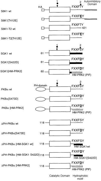
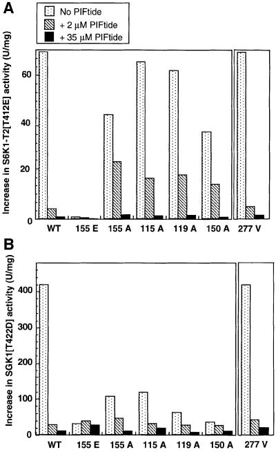
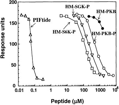
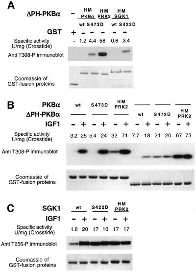
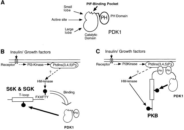
PKB/Akt, S6K1 and SGK are related protein kinases activated in a PI 3-kinase-dependent manner in response to insulin/growth factors signalling. Activation entails phosphorylation of these kinases at two residues, the T-loop and the hydrophobic motif. PDK1 activates S6K, SGK and PKB isoforms by phosphorylating these kinases at their T-loop. We demonstrate that a pocket in the kinase domain of PDK1, termed the 'PIF-binding pocket', plays a key role in mediating the interaction and phosphorylation of S6K1 and SGK1 at their T-loop motif by PDK1. Our data indicate that prior phosphorylation of S6K1 and SGK1 at their hydrophobic motif promotes their interaction with the PIF-binding pocket of PDK1 and their T-loop phosphorylation. Thus, the hydrophobic motif phosphorylation of S6K and SGK converts them into substrates that can be activated by PDK1. In contrast, the PIF-binding pocket of PDK1 is not required for the phosphorylation of PKBalpha by PDK1. The PIF-binding pocket represents a substrate recognition site on a protein kinase that is only required for the phosphorylation of a subset of its physiological substrates.


Figures

None
Fig. 1. The wild-type and mutant AGC kinases used in this study. The circle indicates the position of the T-loop phosphorylation site and the triangle marks the position of the hydrophobic motif phosphorylation site. wt indicates wild type. The C-terminal 104 residues of S6K1 encompass an autoinhibitory domain and this is deleted in the S6K1-T2 construct. The SGK1 protein expressed in this study lacks the 60 N-terminal amino acids. ΔPH-PKBα is a mutant of PKBα that lacks the PH domain. ΔPH-PKBα[HM-SGK1 wt] comprises residues 118–439 of PKBα fused to residues 288–431 of human SGK1. PKBα[HM-SGK1 S422D] comprises residues 118–439 of PKBα fused to residues 288–431 of human SGK1 in which the hydrophobic motif phosphorylation site Ser422 is changed to Asp. PKBα[HM-PRK2] and SGK1[HM-PRK2] comprise residues 118–425 of PKBα and residues 60–372 of SGK1, respectively, fused to residues 934–984 of human PRK2.
None
Fig. 2. Role of the PIF-binding pocket of PDK1 in the phosphorylation of S6K1, SGK1 and PKBα. The indicated AGC substrates were incubated with wild-type GST–PDK1 or mutant GST–PDK1[L155E] in the presence of magnesium and [γ-32P]ATP as described in the Materials and methods. The activation of each AGC kinase was assessed by its ability to phosphorylate the peptide substrate Crosstide (GRPRTSSFAEG). Phosphorylation of the AGC kinase substrate was determined following electrophoresis on a 4–12% gradient polyacrylamide gel and the Coomassie Blue-staining bands corresponding to each substrate were analysed on a phosphoimager. Phosphorylation was also analysed by subjecting the AGC kinases to immuno blotting with antibodies that specifically detect the T-loop-phosphorylated form of S6K1 (Thr252), SGK1 (Thr256) and PKBα (Thr308). Under the conditions used, phosphorylation of each substrate by PDK1 was linear with time and with the amount of enzyme added to the assay. Experiments were performed in duplicate using at least two separate time points. Duplicates within one experiment did not vary >10%, and usually <5%.
None
Fig. 3. Effect of PIFtide on the ability of PDK1 to phosphorylate S6K1, SGK1 and PKBα. The indicated AGC kinase substrates were phosphorylated with wild-type PDK1 in the absence (–) or presence (+) of 2 µM PIFtide in the presence of magnesium and [γ-32P]ATP as described in the legend to Figure 2.
None
Fig. 4. Further evidence that the PIF-binding pocket of PDK1 is required for PDK1 to activate S6K1 and SGK1. S6K1-T2[T412E] (A) and SGK1[S422D] (B) were phosphorylated with the indicated mutants of PDK1 in the absence (dotted bars) or presence of 2 µM PIFtide (hatched bars) or 35 µM PIFtide (filled bars) in the presence of magnesium and [γ-32P]ATP as described in the Materials and methods. The activation of each substrate was assessed by its ability to phosphorylate the peptide substrate Crosstide (GRPRTSSFAEG). The results are presented ±SEM for two separate experiments; each determination was carried out in duplicate.
None
Fig. 5. Interaction of S6K1 and SGK1 with PDK1. (A) 293 cells were transiently transfected with DNA constructs expressing GST, wild type (wt) GST-PDK1 or mutant GST–PDK1[L155E] together with the indicated wild type and mutant forms of HA-epitope-tagged S6K1. Thirty-six hours post-transfection the cells were lysed and the GST fusion proteins were purified by affinity chromatography on glutathione–Sepharose beads. Aliquots of the purified protein were electrophoresed on a 10% SDS–polyacrylamide gel, and immunoblotted using an anti-HA antibody to detect HA-S6K1 or Coomassie to ensure similar expression of GST fusion proteins. To establish that the wild-type and mutant S6K1 forms were expressed at similar levels, 10 µg of total 293 cell lysate (termed Crude extract) for each condition were electrophoresed on a 10% SDS–polyacrylamide gel and immunoblotted with anti-HA antibodies. (B) As above except that 293 cells were transiently transfected with DNA constructs expressing Myc-PDK1 and GST–SGK1 or GST–SGK1[S422D]. Detection of PDK1 was performed by immunoblotting with anti-Myc-antibodies. Duplicates of each condition are shown. Similar results were obtained in two different experiments.
None
Fig. 6. Relative affinities of peptides derived from the hydrophobic motif of S6K1, SGK1, PKBα and PRK2. Surface plasmon resonance measurements were carried out on a BIAcore instrument as described in Materials and methods. Biotinylated PIFtide was immobilized on a strepavidin-coated sensor chip and the specific interaction with GST–PDK1 was recorded in the absence or presence of the indicated concentrations of peptides that encompass the hydrophobic motif (HM) of PRK2, S6K1, SGK1 and PKBα. The peptide PIFtide (open triangles) corresponds to residues 957–980 of PRK2; the peptide HM-S6K1-P (open squares) corresponds to residues 401–418 of S6K1 in which Thr412 is phosphorylated; the peptide HM-SGK1-P (inverted triangles) corresponds to residues 411–428 of SGK1 in which Ser422 is phosphorylated; the peptide HM-PKBα-P (open circles) corresponds to residues 461–480 of PKBα in which Ser473 is phosphorylated; the peptide HM-PKBα (closed circles) corresponds to the non-phosphorylated peptide comprising residues 461–480 of PKBα. The PRK2-derived peptide PIFtide was previously characterized to specifically interact with the PIF-binding pocket on PDK1 (Biondi et al., 2000). Data are single determinations from a representative experiment that was repeated at least twice with similar results.
None
Fig. 7. Effect of swapping the hydrophobic motif of AGC kinases. GST fusion proteins coding for indicated wild-type and mutant forms of PKBα (A and B) and SGK1 (C), whose structure is described in Figure 1, were expressed in 293 cells. The cells were deprived of serum overnight and either left unstimulated or stimulated with 100 ng/ml IGF1 for either 10 min (B) or 15 min (C). The cells were lysed and the GST fusion proteins affinity purified on glutathione–Sepharose. A 0.5 µg sample of each protein was electrophoresed on a 4–12% SDS–polyacrylamide gel, and immunoblotted using a phospho-specific antibody recognizing PKBα (A and B) or SGK1 (C) phosphorylated at their T-loop residue, namely Thr308 and Thr256, respectively. The gels were also stained with Coomassie to ensure similar loading of the GST-ΔPH-PKBα fusion proteins. The specific activity of each GST fusion protein kinase (50 ng) was assessed by its ability to phosphorylate the peptide substrate Crosstide (GRPRTSSFAEG) as described in Materials and methods. The results of a single experiment are shown, similar results were obtained in three separate experiments for (A) and two experiments for (B and C).
None
Fig. 8. Model by which PDK1 specifically recognizes S6K, SGK and PKB. (A) Domain structure of PDK1 indicating where the PIF-binding pocket is located on the small lobe of the kinase domain. (B) Summary of the model by which PDK1 can recognize, interact and then phosphorylate S6K and SGK. In this model PtdIns(3,4,5)P3 regulates the activity of an unidentified hydrophobic motif (HM) kinase that phosphorylates S6K1 and SGK1, thus triggering the docking to the PIF-binding pocket of PDK1. (C) In contrast, the PIF-binding pocket of PDK1 is not involved in the binding of PDK1 to PKB. Instead it is the interaction of the PH domains of PKB and PDK1 with PtdIns(3,4,5)P3 that brings PKB and PDK1 together.

References

    1. Alessi D.R., Cohen,P., Ashworth,A., Cowley,S., Leevers,S.J. and Marshall,C.J. (1995) Assay and expression of mitogen-activated protein kinase, MAP kinase kinase and Raf. Methods Enzymol., 255, 279–290. - PubMed
    1. Alessi D.R., Deak,M., Casamayor,A. et al. (1997a) 3-phosphoinositide-dependent protein kinase-1 (PDK1): structural and functional homology with the Drosophila DSTPK61 kinase. Curr. Biol., 7, 776–789. - PubMed
    1. Alessi D.R., James,S.R., Downes,C.P., Holmes,A.B., Gaffney,P.R., Reese,C.B. and Cohen,P. (1997b) Characterization of a 3-phospho inositide-dependent protein kinase which phosphorylates and activates protein kinase Bα. Curr. Biol., 7, 261–269. - PubMed
    1. Alessi D.R., Kozlowski,M.T., Weng,Q.P., Morrice,N. and Avruch,J. (1998) 3-phosphoinositide-dependent protein kinase 1 (PDK1) phosphorylates and activates the p70 S6 kinase in vivo and in vitro. Curr. Biol., 8, 69–81. - PubMed
    1. Andjelkovic M., Alessi,D.R., Meier,R. et al. (1997) Role of translocation in the activation and function of protein kinase B. J. Biol. Chem., 272, 31515–31524. - PubMed

Publication types

MeSH terms