. 2001 Aug 15;20(16):4443-53.
doi: 10.1093/emboj/20.16.4443.

HBV infection of cell culture: evidence for multivalent and cooperative attachment

Affiliations

HBV infection of cell culture: evidence for multivalent and cooperative attachment

N Paran et al. EMBO J. .

Abstract

Hepadnaviruses do not infect cultured cells, therefore our knowledge of the mechanism of the early stages of virus-cell interaction is rather poor. In this study, we show that dimethylsulfoxide (DMSO)-treated HepG2 hepatoblastoma cells are infected efficiently by serum-derived hepatitis B virus (HBV) as monitored by viral gene expression and replication markers. To measure virus attachment, a variety of HBV surface proteins (HBsAgs) were conjugated to polystyrene beads and their capacity to attach cells was visualized and quantified by light microscopy at a single-cell resolution. Remarkably, DMSO increases the attachment efficiency by >200-fold. We further identify the QLDPAF sequence within preS1 as the receptor-binding viral domain epitope. Interestingly, a similar sequence is shared by several cellular, bacterial and viral proteins involved in cell adhesion, attachment and fusion. We also found that the small HBsAg contains a secondary attachment site that recognizes a distinct receptor on the cell membrane. Furthermore, we provide evidence in support of multivalent HBV attachment with synergistic interplay. Our data depict a mechanistic view of virus attachment and ingestion.


Figures
None
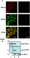
Fig. 1. Efficient infection of DMSO-treated HepG2 cells by HBV. (A) HepG2 cells were seeded on 18 mm coverslips and either DMSO treated or left untreated. After 6 days, cells were incubated for 14 h with HBV-positive sera containing 109 particles per ml, diluted in culture media. Subsequently, the unbound viruses were discarded and the cells were incubated further with (left panel) or without (right panel) DMSO for an additional 4 days. Viral infection was monitored by indirect immunoflourescent staining with αHBcAg (red) and αHBsAg (green) polyclonal sera, and by either RRX- or FITC-conjugated antibodies, respectively. The stained cells were visualized by scanning laser confocal microscopy. Yellow represents co-localization of HBcAg and αHBsAg. (B) Viral DNA in the duplicate samples was isolated from the infected cells and from the virions and subjected to PCR to detect the presence of HBV RC and cccDNA. The PCR results of the viral sample (lane 1), and extracts of infected cells that were DMSO treated (lane 2) or left untreated (lane 3) are shown. Control PCRs were performed for the endogenous AML-2. The migration position of the DNA is shown as the molecular weight in kb. (C) Southern blot analysis of total (lanes 2–9) and extrachromosomal DNA (lanes 10–11), extracted from HBV- and mock-infected (lane M) cells at the indicated days post-infection (dpi). Cells were either DMSO treated (lanes 2, 5–9 and 11) or left untreated (lanes 3, 4 and 10). The gel migration positions of relaxed circular (RC), covalently closed circular (ccc) and single-stranded (ss) forms are indicated. In lane 8, 100 µM lamivudine (3TC) was added to the culture medium 14 h after infection. Fresh medium containing 3TC was added every 3 days. To block infection by MA 18/7 neutralizing monoclonal anti-preS1 antibody, 200 µl of HBV-positive serum was pre-incubated with 0.5 µg of IgG for 4 h before infection. MW = DNA molecular weight in kb.
None
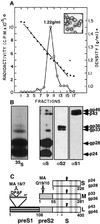
Fig. 2. Recombinant HBV SVPs containing all the HBV surface proteins. CHO cells stably transfected with the AL26 plasmid express 22 nm HBV subparticles. (A) Purified particles were fractionated on a CsCl gradient and the resulting fractions were assayed for HBsAg by radioimmunoassay using 125I-labeled anti-HBsAg antibodies. The density of HBsAg SVPs was calculated to be 1.22 g/ml, characteristic of the 22 nm spherical HBsAg SVPs. The insert shows 22 nm particles that were negatively stained by uranyl acetate followed by TEM. (B) The composition of the various surface proteins was determined by [35S]methionine labeling on SDS–PAGE and by western analysis with MA 18/7, the antibody specific for preS1 (αS1), MA Q19/10 (for preS2), and with polyclonal anti-S antibodies (αS). The various HBV surface proteins are indicated. (C) Schematic presentation of the different (S, L and M) HBsAg proteins with the corresponding antibody epitopes. The gray boxes represent the transmembrane regions.
None
Fig. 3. DMSO improves SVPs attachment. (A) HepG2 cells were seeded on coverslips and incubated with beads coated with either recombinant SVPs or BSA. Non-attached beads were removed and the cells were fixed and visualized by DIC light microscopy at 66× magnification. Attachment of beads was quantified for each representative microscopic field and the average attachment for a given number of beads per cell was determined. (B) The percentage of cells that attached beads out of the overall cell population (total) and the percentage of cells out of the total population that attached more than four beads per cell (high) for either BSA– or SVP–beads are presented together with their respective standard deviation bars. (C and D) As in (A) and (B), but cells were treated with 2% DMSO.
None
Fig. 4. Attachment of SVP-conjugated beads is accompanied by endocytosis. (AD) Late stages (A, B and D) and completion of SVP–bead engulfment (C) by the cell membrane are visualized by TEM. (B) is a higher magnification of (A). (E–H) Poor (E) and efficient (F) endocytosis of SVP–beads by untreated and DMSO-treated HepG2 cells, respectively, as visualized by SEM. Various stages of bead internalization are demonstrated (G and H).
None
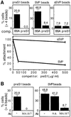
Fig. 5. The preS1 epitope (amino acids 21–47) mediates cell attachment. (A) Beads conjugated with either BSA, SVPs, recombinant preS1 or synthetic peptide encompassing amino acids 21–47 of preS1 were incubated with DMSO-treated cells. The percentage of cells out of the total population that attached more than four beads per cell was calculated and is presented together with their respective standard deviation bars. (B) The experiment described in (A) was repeated with untreated HepG2 cells.
None
Fig. 6. The QLDPAF motif in preS1 mediates preS1 attachment. (A) Sequence comparison of the wild-type QLDPAF preS1 and of the scrambled mutant protein. (B) Preferential detection of wild-type versus mutant preS1 by anti-preS1 (MA 18/7) (upper panel); there was equal detection by α6His. (C) Attachment efficiencies of beads conjugated to the indicated proteins in the presence or absence of pre-incubated mutant preS1 as a competitor. The attachment assay and the quantification of attached beads were performed as described in Figure 2. (D) Database analysis revealed that the QLDPA sequence is also found in pX, a second HBV protein. Also, a sequence similar to this motif was found in a number of viral bacterial and cellular proteins that are involved in cell adhesion, attachment and fusion.
None
Fig. 7. Small SVPs (sSVPs) lacking the preS1 region contain an independent attachment region. (A) Recombinant sSVPs were produced by transfection of HEK 293 cells with the pMH8 plasmid. Western analysis with polyclonal anti-HBsAg (αS) and monoclonal anti-preS1 antibodies (MA 18/7) was performed to compare the composition of the various HBsAgs in sSVPs versus the recombinant SVPs. The various HBsAgs are indicated. (B) Both SVPs and the sSVPs were conjugated to beads and their ability to mediate bead attachment in the presence or absence of DMSO was determined. See Figure 2 for the structure of the different HBsAg proteins.
None
Fig. 8. Cooperative action of multivallent attachment sites in SVPs. (A) DMSO-treated HepG2 cells were pre-incubated for 2 h with 500 µg/ml of either BSA or recombinant preS1 as competitors prior to addition of preS1-conjugated beads. The soluble competitor proteins were pre-incubated at 100× the concentration of the conjugated ligand. The percentage of cells of the total population that attached more than four beads per cell is shown (upper panels). The experiment was carried out with an increasing amount of the competitor as indicated (lower panel). (B) The attachment of the preS1–beads was neutralized by pre-incubation of the beads for 2 h with either monoclonal anti-preS1 (MA 18/7) or the control monoclonal anti-HBcAg (ns, non-specific), as indicated.

Similar articles

Cited by

References

    1. Breiner K.M., Urban,S. and Schaller,H. (1998) Carboxypeptidase D (gp180), a Golgi-resident protein, functions in the attachment and entry of avian hepatitis B viruses. J. Virol., 72, 8098–8104. - PMC - PubMed
    1. Bruns M., Miska,S., Chassot,S. and Will,H. (1998) Enhancement of hepatitis B virus infection by noninfectious subviral particles. J. Virol., 72, 1462–1468. - PMC - PubMed
    1. De Meyer S., Gong,Z.J., Suwandhi,W., van Pelt,J., Soumillion,A. and Yap,S.H. (1997) Organ and species specificity of hepatitis B virus (HBV) infection: a review of literature with a special reference to preferential attachment of HBV to human hepatocytes. J. Viral Hepat., 4, 145–153. - PubMed
    1. Faktor O., De-Medina,T. and Shaul,Y. (1988) Regulation of hepatitis B virus S gene promoter in transfected cell lines. Virology, 162, 362–368. - PubMed
    1. Gripon P., Diot,C., Corlu,A. and Guguen-Guillouzo,C. (1989) Regulation by dimethylsulfoxide, insulin and corticosteroids of hepatitis B virus replication in a transfected human hepatoma cell line. J. Med. Virol., 28, 193–199. - PubMed

Publication types

MeSH terms

Substances