. 2001 Aug 15;20(16):4454-66.
doi: 10.1093/emboj/20.16.4454.

Hes1 and Hes3 regulate maintenance of the isthmic organizer and development of the mid/hindbrain

Affiliations

Hes1 and Hes3 regulate maintenance of the isthmic organizer and development of the mid/hindbrain

H Hirata et al. EMBO J. .

Abstract

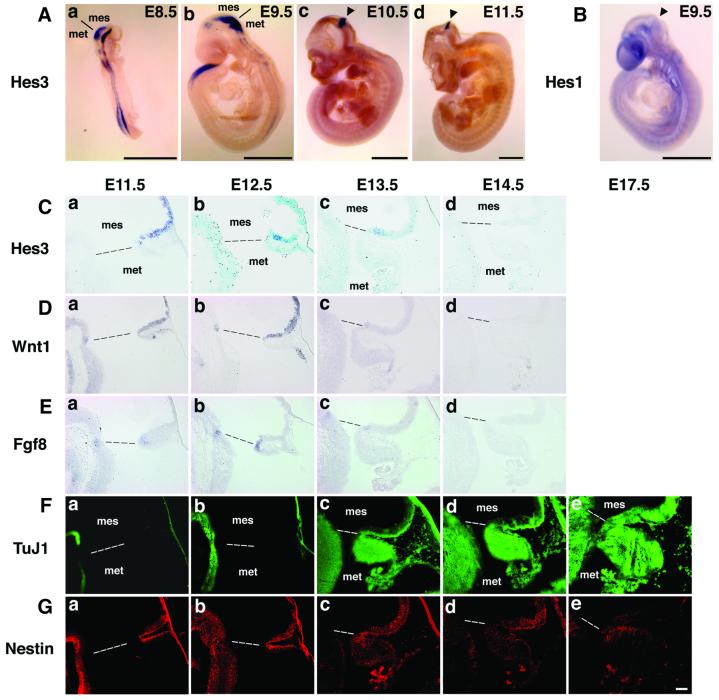
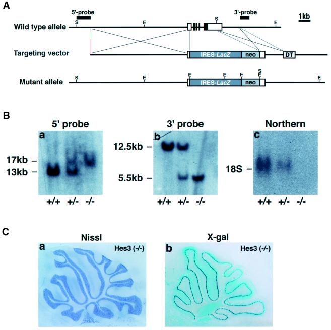
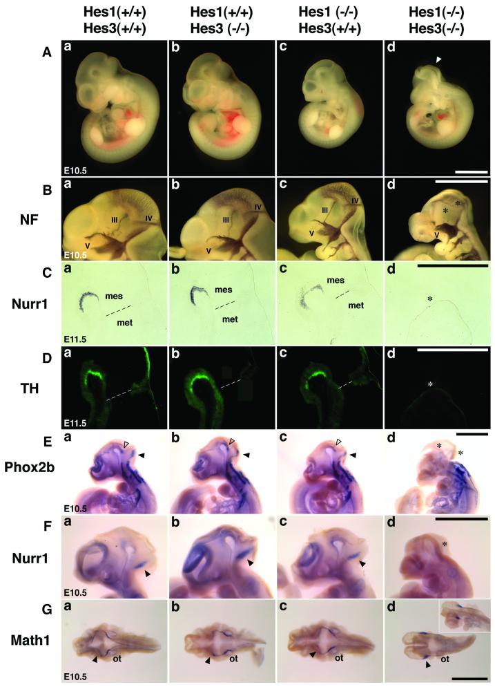
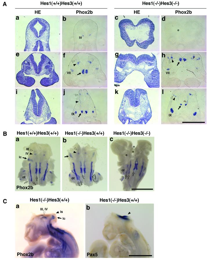
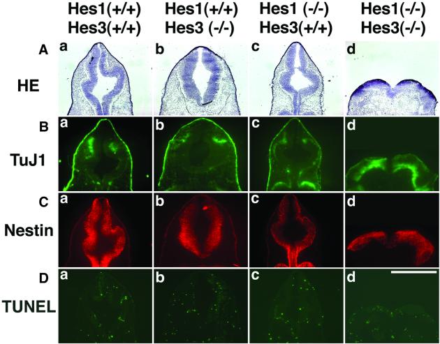
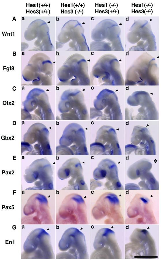
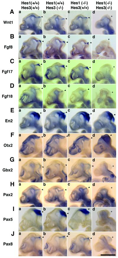
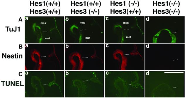
The isthmic organizer, which is located at the midbrain-hindbrain boundary, plays an essential role in development of the midbrain and anterior hindbrain. It has been shown that homeobox genes regulate establishment of the isthmic organizer, but the mechanism by which the organizer is maintained is not well understood. Here, we found that, in mice doubly mutant for the basic helix-loop-helix genes Hes1 and Hes3, the midbrain and anterior hindbrain structures are missing without any significant cell death. In these mutants, the isthmic organizer cells prematurely differentiate into neurons and terminate expression of secreting molecules such as Fgf8 and Wnt1 and the paired box genes Pax2/5, all of which are essential for the isthmic organizer function. These results indicate that Hes1 and Hes3 prevent premature differentiation and maintain the organizer activity of the isthmic cells, thereby regulating the development of the midbrain and anterior hindbrain.


Figures

None
Fig. 1. Spatio-temporal expression of Hes3. (A) In situ hybridization of Hes3. (a) At E8.5, Hes3 is expressed widely in the midbrain–hindbrain region and spinal cord but not in the forebrain. (b) At E9.5, Hes3 expression in the spinal cord is down-regulated. (c and d) At E10.5 and E11.5, Hes3 expression is restricted to the midbrain–hindbrain boundary region. The isthmic organizer is indicated by arrowheads. (B) In situ hybridization of Hes1. At E9.5, Hes1 is also expressed in the midbrain–hindbrain boundary region (arrowhead). (C–G) In situ hybridization (C–E) and immuno histochemistry (F and G) of parasagittal sections. In all sections, ventral is to the left. (C) (a–c) Hes3 is expressed in the ventricular zone of the rostral side of the midbrain–hindbrain boundary at E11.5–13.5. (d) At E14.5, Hes3 expression almost disappears. (D) (a and b) At E11.5–12.5, Wnt1 is expressed in the ventricular zone of the rostral side of the midbrain–hindbrain boundary. (c and d) Wnt1 expression is down-regulated at E13.5 and disappears at E14.5. (E) (a and b) At E11.5–12.5, Fgf8 is expressed in the ventricular zone of the caudal side of the midbrain–hindbrain boundary. (c and d) Fgf8 expression is down-regulated at E13.5 and disappears at E14.5. (F) Immunohistochemistry of TuJ1. (a) At E11.5, some TuJ1+ neurons were differentiated in the ventral side of the midbrain and hindbrain but not in the isthmic region. (b) At E12.5, some TuJ1+ neurons were differentiated in the ventral side of the isthmic region. (c and d) At E13.5–14.5, some TuJ1+ neurons were also differentiated in the dorsal side of the isthmic region. (e) At E17.5, almost all cells differentiate into neurons. (G) Immunohistochemistry of Nestin. (a–d) At E11.5–13.5, Nestin+ neural precursor cells are present in the ventricular zone of the isthmic region but significantly decreased at E14.5. (e) At E17.5, there are virtually no neural precursor cells left in the isthmic region. mes, mesencephalon; met, metencephalon. Scale bar, 1 mm (A and B); 100 µm (C–G).
None
Fig. 2. Targeted deletion of Hes3. (A) Genomic structure of mouse Hes3 and targeting strategy. The coding and non-coding regions are indicated by closed and open boxes, respectively. All the coding region is replaced with lacZ-neo. lacZ expression is under the control of the Purkinje cell-specific promoter (Hirata et al., 2000). E, EcoRI; S, SalI. (B) Southern and northern analyses. (a and b) Genotypes were determined by Southern blot analysis. Genomic DNA was digested with SalI (a) and EcoRI (b). (c) Northern analysis shows that Hes3 expression is decreased in heterozygous and abolished in homozygous embryos. (C) Histochemistry of Hes3(–/–) mice. (a) Nissl staining. (b) X-gal staining. Purkinje cells (X-gal+) are generated normally in Hes3(–/–) cerebellum.
None
Fig. 3. Defects of midbrain and anterior hindbrain patterning of Hes1–Hes3 double-mutant embryos. (A) E10.5 embryos. The Hes1(–/–)–Hes3(–/–) embryo displays the neural tube defect (d, arrowhead). (B) Whole-mount immunostaining with anti-neurofilament (NF) antibody of E10.5 embryos. Cranial nerves III and IV are generated in the wild-type, Hes1(–/–) and Hes3(–/–) embryos whereas they are missing in the Hes1(–/–)–Hes3(–/–) embryo (d, asterisks), suggesting that midbrain development is affected in the double-mutant embryos. Cranial nerve V is generated normally in the double mutant. (CNurr1 expression in the ventral midbrain at E11.5. This expression domain is missing in the Hes1–Hes3 double-mutant embryo (d, asterisk). (D) Immunohistochemistry with anti-tyrosine hydroxylase (TH) antibody. Dopaminergic neurons (TH+) are missing in the ventral midbrain of the Hes1–Hes3 double-mutant embryo at E11.5 (d, asterisk). (EPhox2b is expressed in oculomotor (III) and trochlear (IV) motor nuclei in the ventral midbrain and isthmus, respectively (a–c, open arrowheads), and in locus ceruleus in rhombomere 1 (a–c, closed arrowheads) of the E10.5 wild-type, Hes1(–/–) and Hes3(–/–) embryos. In contrast, such neurons are missing in the Hes1–Hes3 double-mutant embryo (d, asterisks). However, Phox2b expression domains in the mid/posterior hindbrain are normal in the Hes1–Hes3 double-mutant embryo. (F) The Nurr1 expression domain observed in the anterior hindbrain of the wild-type, Hes1(–/–) and Hes3(–/–) embryos (a–c, arrowheads) is missing in the Hes1–Hes3 double-mutant embryo at E10.5 (d, asterisk). (GMath1 is expressed in the rhombic lip (arrowheads) of the wild-type, Hes1(–/–), Hes3(–/–) and Hes1(–/–)–Hes3(–/–) double-mutant embryos at E10.5. Note that the rhombic lips are distorted due to the neural tube defect in the double mutant (d). mes, mesencephalon; met, metencephalon; ot, otic vesicle. Scale bar, 1 mm.
None
Fig. 4. Patterning defects of the midbrain and anterior hindbrain are specific to Hes1–Hes3 double-mutant embryos. (A) HE staining and in situ hybridization for Phox2b expression with frontal sections of E10.5 embryos. Trochlear motor nuclei (IV) are missing in the double-mutant isthmus region (d, asterisk). In contrast, three columns of the Phox2b expression domains are generated in the double-mutant hindbrain (h and l, arrows and arrowheads). Cranial nerves VII and IX are also present in the double-mutant embryos (h and l). In all sections, dorsal is up. (B) Flat-mount in situ hybridization of E10.5 wild-type (a), Hes1(–/–) (b) and Hes1(–/–)–Hes3(–/–) (c) embryos. The dorsal side is excised. In Hes1(–/–) embryo (b), which has neural tube defects, oculomotor (open arrowhead) and trochlear (closed arrowhead) motor nuclei and locus ceruleus (arrow) are generated. In contrast, in the double mutant (c), those structures are missing (asterisks). However, the three columns of Phox2b expression domains are present in the double-mutant hindbrain (open arrows). ot, otic vesicle. (C) Whole-mount in situ hybridization of E10.5 Hes1(–/–) embryos with neural tube defects. (a) The Phox2b expression domains (oculomotor and trochlear motor nuclei and locus ceruleus) are present, indicating that neural tube defects do not cause the lack of the midbrain and anterior hindbrain structures at this stage. III, oculomotor; IV, trochlear; is, isthmus; lc, locus ceruleus. (b) Pax5 is expressed in the midbrain–hindbrain boundary region, indicating that neural tube defects do not cause the lack of the isthmic organizer at this stage. The isthmus is indicated by an arrowhead. Scale bar, 1 mm.
None
Fig. 5. Premature neuronal differentiation in the Hes1–Hes3 double-mutant midbrain. (A) HE staining of frontal sections of midbrain at E10.5. (B and C) Frontal sections of midbrain at E10.5 were subjected to immunohistochemistry with TuJ1 (B) and anti-Nestin antibodies (C). In the wild-type, Hes1(–/–) and Hes3(–/–) midbrain, some neurons (TuJ1+) are generated (Ba–c). In Hes1(–/–)–Hes3(–/–) midbrain, more neurons are generated prematurely (Bd), suggesting that Hes1 and Hes3 cooperatively prevent premature neurogenesis. In spite of this accelerated neurogenesis, there are still abundant neural precursor cells (Nestin+) in the double-mutant midbrain (Cd). (D) TUNEL analysis of frontal sections of midbrain at E10.5. No significant difference is observed in the double mutant, indicating that cell death is not induced by Hes1–Hes3 double mutation. Note that the double-mutant midbrain displays an exencephaly. In all sections, dorsal is up. Scale bar, 1 mm.
None
Fig. 6. The isthmic organizer is established in Hes1–Hes3 double-mutant embryos at E9.5. Whole-mount in situ hybridization of E9.5 embryos. (ADWnt1, Fgf8, Otx2 and Gbx2 are expressed normally in the double-mutant embryos (arrowheads). (E) The paired box gene Pax2 is expressed in the wild-type, Hes1(–/–) and Hes3(–/–) isthmic organizer (a–c, arrowheads). In contrast, the expression is missing in the double mutant (d, asterisk). (F and GPax5 and En1 expression is not significantly changed in the double mutant although the expression domain is somewhat distorted by the neural tube defect (d, arrowhead). Scale bar, 1 mm.
None
Fig. 7. The isthmic organizer is not maintained properly in Hes1–Hes3 double-mutant embryos at E10.5. (AWnt1 is expressed in the wild-type, Hes1(–/–) and Hes3(–/–) isthmic organizer (a–c, arrowheads) whereas the expression disappears prematurely in the double mutant at E10.5 (d, asterisk). (BDFgf8/17/18 are expressed in the wild-type, Hes1(–/–) and Hes3(–/–) isthmic organizer (a–c, arrowheads) whereas the expression is not detectable in the double mutant at E10.5 (d, asterisks). Note that Fgf8/17 expression in the forebrain is not affected in the double mutants (Bd and Cd). (EEn2 is expressed normally in the midbrain–hindbrain region of the double mutant (arrowhead). (FOtx2 expression is restricted to the region rostral to the isthmus of the wild-type, Hes1(–/–) and Hes3(–/–) embryos (a–c). In contrast, Otx2 expression is extended caudally in the double mutant (d). The isthmus is indicated by an arrowhead. (GGbx2 is expressed in the hindbrain of the wild-type, Hes1(–/–) and Hes3(–/–) embryos (a–c). In contrast, Gbx2 expression is not detectable in the anterior hindbrain of the double mutant (d, asterisk). The isthmus is indicated by an arrowhead. (HJPax2/5/8 are expressed in the wild-type, Hes1(–/–) and Hes3(–/–) isthmic organizer (a–c, arrowheads). In contrast, the expression of Pax2 and Pax5 is missing and that of Pax8 is significantly down-regulated in the double mutant (d, asterisks). Thus, in the absence of Hes1 and Hes3, the isthmic organizer is not maintained properly. The staining of Pax2 in the floor plate (H) is an artifact. Scale bar, 1 mm.
None
Fig. 8. Premature neuronal differentiation in the Hes1–Hes3 double-mutant isthmic organizer. Parasagittal sections of E11.5 embryos were examined by immunohistochemistry. (A) (a–c) TuJ1+ neurons are not generated in the wild-type, Hes1(–/–) and Hes3(–/–) isthmic region whereas some neurons are already differentiated outside this region. (d) Almost all cells prematurely differentiate into neurons in the double-mutant isthmic organizer. (B) (a–c) There are many Nestin+ neural precursor cells in the wild-type, Hes1(–/–), and Hes3(–/–) isthmic region. (d) Nestin+ neural precursor cells are virtually absent from the double-mutant isthmus and the surrounding region. (C) TUNEL assay. TUNEL+ cells are not increased in the double-mutant isthmic organizer. Scale bar, 1 mm.

References

    1. Akazawa C., Ishibashi,M., Shimizu,C., Nakanishi,S. and Kageyama,R. (1995) A mammalian helix–loop–helix factor structurally related to the product of Drosophila proneural gene atonal is a positive transcriptional regulator expressed in the developing nervous system. J. Biol. Chem., 270, 8730–8738. - PubMed
    1. Allen T. and Lobe,C.G. (1999) A comparison of Notch, Hes and Grg expression during murine embryonic and post-natal development. Cell. Mol. Biol., 45, 687–708. - PubMed
    1. Bally-Cuif L., Cholley,B. and Wassef,M. (1995) Involvement of Wnt1 in the formation of the mes/metencephalic boundary. Mech. Dev., 53, 23–34. - PubMed
    1. Beddington R.S.P. and Robertson,E.J. (1998) Anterior patterning in mouse. Trends Genet., 14, 277–284. - PubMed
    1. Ben-Arie N., Hassan,B.A., Bermingham,N.A., Malicki,D.M., Armstrong,D., Matzuk,M., Bellen,H.J. and Zoghbi,H.Y. (2000) Functional conservation of atonal and Math1 in the CNS and PNS. Development, 127, 1039–1048. - PubMed

Publication types

MeSH terms