. 2001 Aug 15;20(16):4478-89.
doi: 10.1093/emboj/20.16.4478.

FANCC interacts with Hsp70 to protect hematopoietic cells from IFN-gamma/TNF-alpha-mediated cytotoxicity

Affiliations

FANCC interacts with Hsp70 to protect hematopoietic cells from IFN-gamma/TNF-alpha-mediated cytotoxicity

Q Pang et al. EMBO J. .

Abstract

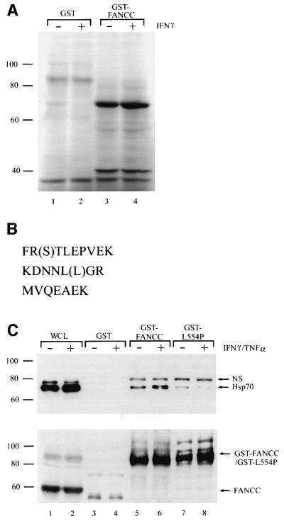
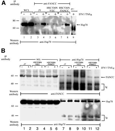
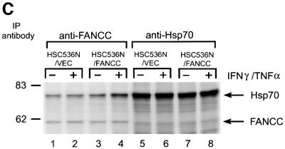
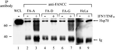
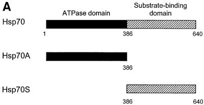
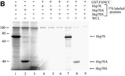
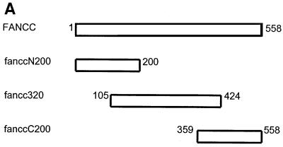
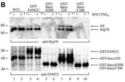
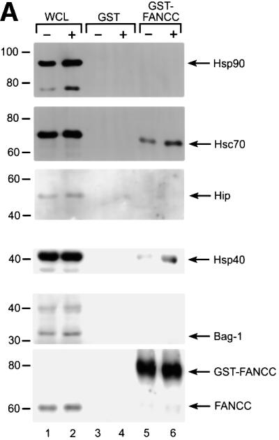
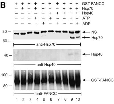
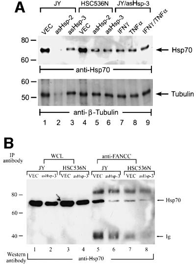
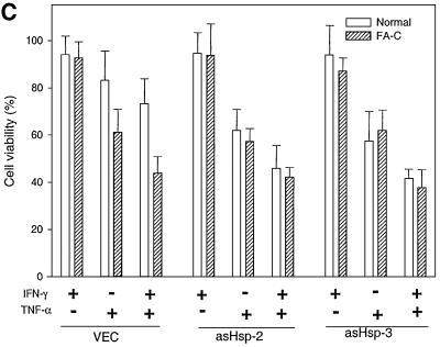
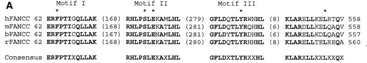
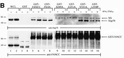
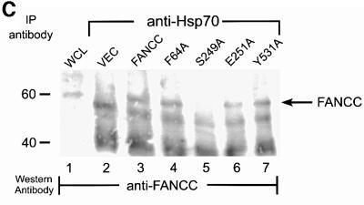
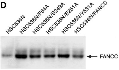
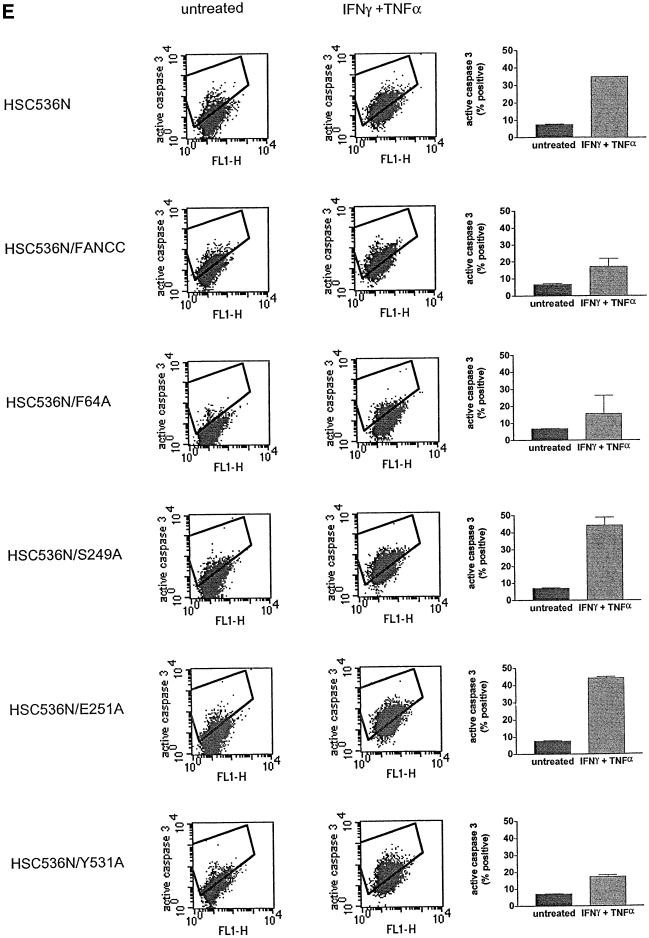
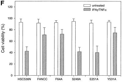
The Fanconi anemia (FA) complementation group C gene product (FANCC) functions to protect hematopoietic cells from cytotoxicity induced by interferon-gamma (IFN-gamma), tumor necrosis factor-alpha (TNF-alpha) and double-stranded RNA (dsRNA). Because apoptotic responses of mutant FA-C cells involve activation of interferon-inducible, dsRNA-dependent protein kinase PKR, we sought to identify FANCC-binding cofactors that may modulate PKR activation. We identified the molecular chaperone Hsp70 as an interacting partner of FANCC in lymphoblasts and HeLa cells using 'pull-down' and co-immunoprecipitation experiments. In vitro binding assays showed that the association of FANCC and Hsp70 involves the ATPase domain of Hsp70 and the central 320 residues of FANCC, and that both Hsp40 and ATP/ADP are required. In whole cells, Hsp70-FANCC binding and protection from IFN-gamma/TNF-alpha-induced cytotoxicity were blocked by alanine mutations located in a conserved motif within the Hsp70-interacting domain of FANCC. We therefore conclude that FANCC acts in concert with Hsp70 to prevent apoptosis in hematopoietic cells exposed to IFN-gamma and TNF-alpha.


Figures

None
Fig. 1. Identification of Hsp70 as an FANCC-interacting protein. (A) Proteins binding to GST–FANCC. GST alone or GST-fused wild-type FANCC (GST–FANCC) was incubated with 35S-labeled whole-cell lysates (WCLs) from JY lymphoblasts treated (+) or not (–) with IFN-γ (10 ng/ml) for 2 h. Bound proteins were analyzed by SDS–PAGE and autoradiography. (B) Amino acid sequences of three peptides derived from microsequencing of the 70 kDa protein. Parentheses indicate probable but not definite residues. (C) Association of FANCC with Hsp70. GST alone, GST–FANCC or GST-fused FA-C patient-derived mutant (GST–L554P) was incubated with WCLs from JY lymphoblasts treated (+) or not (–) with IFN-γ and TNF-α (10 ng/ml each), and the bound proteins were analyzed by immuno blotting with anti-Hsp70 antibody. The same WCL (100 µg) was electrophoresed directly as input control. NS, non-specific.
None
Fig. 2. Co-immunoprecipitation of endogenous FANCC and Hsp70. (A) Normal JY and FA-C HSC536N lymphoblasts transduced with vector alone (HSC/VEC) or the wild-type FANCC cDNA (HSC/FANCC) were treated (+) or not (–) with IFN-γ and TNF-α (10 ng/ml each) for 2 h. WCLs (1 mg of total proteins) were immunoprecipitated (IP) with a polyclonal FANCC antibody (lanes 3–8) or pre-immune serum (lane 9). The immunocomplexes were then analyzed by western blotting with a monoclonal antibody specific for Hsp70. Lanes 1 and 2 are direct western blotting with 100 µg of WCLs from untreated (–) or treated (+) JY lymphoblasts. (B) WCLs prepared as described in (A) were immunoprecipitated with anti-Hsp70 and probed with anti-FANCC (top) and anti-Hsp70 (bottom). Input controls (lanes 1–6) were analyzed with 100 µg of WCL. (C) Stoichiometry of the FANCC–Hsp70 association. A 200 µg aliquot of 35S-labeled WCL was immunoprecipitated with antibodies for either FANCC or Hsp70. The amounts of Hsp70 co-immunoprecipitated by anti-FANCC (lanes 1–4) were compared with the amounts of total cellular Hsp70 immunoprecipitated directly with anti-Hsp70. The same comparison was applied to the FANCC protein (lanes 5–8).
None
Fig. 3. Immunoprecipitation–western analysis of WCLs from FA-A and FA-D patient-derived lymphocytes, FA-G patient-derived fibroblasts and HeLa cells treated (+) or not (–) with IFN-γ and TNF-α (10 ng/ml each). Equal amounts of WCLs (1 mg of total proteins each) were immunoprecipitated with anti-FANCC, followed by western analysis with anti-Hsp70. A 100 µg aliquot of FA-A WCL was used as an input control (lane 1).
None
Fig. 4. The ATPase domain of Hsp70 interacts with FANCC. (A) A diagram of full-length human Hsp70 (residues 1–640) and its deletion derivatives Hsp70A (residues 1–368 comprised of the ATPase domain) and Hsp70S (residues 386–640 comprised of the substrate-binding domain). (B) Interaction of FANCC with 35S-labeled Hsp70 proteins. GST–FANCC was absorbed to glutathione–Sepharose beads and incubated with 20 µl of [35S]methionine-labeled Hsp70 proteins. Eluted proteins were resolved by SDS–PAGE and analyzed by autoradiography. Lanes 1–3, 5 µl of each 35S-labeled full-length Hsp70, Hsp70A and Hsp70S as input controls; lanes 4–6, GST–FANCC incubated with the indicated 35S-labeled Hsp70 proteins; lanes 7–9, GST–FANCC incubated with the indicated 35S-labeled Hsp70 proteins plus 100 µg of WCL from IFN-γ/TNF-α-treated JY lymphoblasts.
None
Fig. 5. The Hsp70-interacting domain is located in the central region of FANCC. (A) A schematic diagram of full-length and deletion mutants of FANCC. Full-length FANCC and truncated mutants fanccN200, fancc320 and fanccC200 are indicated as the regions of FANCC fused to GST. (B) Interaction of Hsp70 with the central region of FANCC. GST-fused full-length and truncated FANCC proteins were incubated with WCLs from JY lymphoblasts treated (+) or not (–) with IFN-γ and TNF-α (10 ng/ml each). The glutathione–Sepharose affinity precipitates were analyzed by SDS–PAGE followed by immuno blotting with anti-Hsp70 antibody (top) and reprobed with anti-FANCC to verify the input of the GST fusion proteins (bottom).
None
Fig. 6. Association of FANCC with Hsp70 is dependent on Hsp40. (A) Identification of Hsp40 as the factor required for FANCC–Hsp70 association. GST alone or GST–FANCC was incubated with WCLs from JY lymphoblasts treated (+) or not (–) with IFN-γ and TNF-α (10 ng/ml each), and the bound proteins were analyzed by immuno blotting with antibodies against the indicated proteins. (B) Hsp40-dependent association of FANCC with Hsp70. GST–FANCC (120 µM)–Sepharose beads were incubated with Hsp70 and/or Hsp40 (5 µM each) in the presence of ATP or ADP (1 mM each) for 15 min at 30°C. The Sepharose was washed and bound materials were analyzed by immunoblotting with antibodies for Hsp70 (top), Hsp40 (middle) or FANCC (bottom).
None
Fig. 7. Inhibition of Hsp70 expression sensitizes normal but not FA-C lymphoblasts to IFN-γ/TNF-α-mediated cytotoxicity. (A) Antisense Hsp70 constructs reduce Hsp70 expression. WCLs (50 µg of total proteins each) from JY and HSC536N lymphoblasts transduced with vector alone (VEC) or two antisense constructs (asHsp-2 or asHsp-3) was analyzed by immunoblotting with anti-Hsp70 (top) or anti-tubulin (bottom) antibody. The levels of Hsp70 in JY lymphoblasts carrying asHsp-3 treated with IFN-γ, TNF-α or both (10 ng/ml each) are indicated in lanes 7, 8 and 9, respectively. (B) Antisense Hsp70 constructs reduce FANCC–Hsp70 complexes. Equal amounts of WCLs (1 mg of total proteins each) from IFN-γ/TNF-α-treated JY and HSC536N lymphoblasts transduced with vector alone (VEC) or an antisense construct (asHsp-3) were immunoprecipitated with anti-FANCC followed by western analysis with anti-Hsp70. A 100 µg aliquot of the same WCL was run as input control (lanes 1–4). (C) Cell viability of JY (normal) and HSC536N (FA-C) lymphoblasts carrying vector alone (VEC) or antisense Hsp70 constructs (asHsp-2 or asHsp-3), in response to IFN-γ or/and TNF-α treatments. Data represent the means ± standard deviations of triplicate determinations.
None
Fig. 8. Alanine-substituted mutations in the Hsp70-interactive domain of FANCC abolish FANCC–Hsp70 interaction and sensitize cells to IFN-γ and TNF-α. (A) Motifs conserved among FANCC homologs of human (hFANCC), mouse (mFANCC), bovine (bFANCC) and rat (rFANCC). Bold letters correspond to identical amino acids, and ‘X’ to non-conserved residues. The number of residues between the different motifs is indicated in parentheses. The asterisks indicate the positions at which alanine substitutions were made and the location of the FA-C patient-derived mutation L554P. (B) Effect of FANCC mutations on interaction between FANCC and Hsp70. WCL (1 mg of protein) of JY lymphoblasts treated with IFN-γ and TNF-α was incubated with GST fusion proteins bound to glutathione–Sepharose beads. The glutathione–Sepharose affinity precipitates were analyzed by SDS–PAGE followed by immunoblotting. Top: the western blot was probed with antibody to Hsp70; bottom, the same blot was reprobed with anti-FANCC to verify the input of the GST fusion proteins. Lanes 1 and 2 were WCLs (50 µg of total proteins each) to show the effects of IFN-γ and TNF-α on expression of Hsp70 and FANCC. (C) Association of alanine-substituted FANCC with Hsp70 in vivo. WCLs (1 mg of total proteins) from PD4 lymphoblasts transduced with vector alone (VEC), the wild-type FANCC (FANCC) or alanine-substituted FANCC cDNA were immuno precipitated (IP) with anti-Hsp70 antibody (lanes 2–7). The immunocomplexes were then analyzed by western blotting with anti-FANCC antibody. Lanes 1 is direct western blotting with 100 µg of WCL from PD4 lymphoblasts. (D) Expression of alanine-substituted FANCC mutant protein in HSC536N cells. WCLs (100 µg each) were subjected to SDS–PAGE and analyzed by immunoblotting with anti-FANCC antibody. (E) Flow cytometric analysis of fractional apoptotic responses in HSC536N lymphoblasts expressing FANCC mutations. Cells were incubated with IFN-γ (10 ng/ml) for 15 h before treatment with 10 ng/ml of TNF-α for 48 h and harvested for flow cytometric analysis. Treatments of HSC/VEC and HSC/FANCC with camptothecin were used as positive controls (50 mM for 9 h) and induced positivity of 65.1 and 22.5%, respectively. A total of 10 000 events for each sample were analyzed. (F) Cell viability of HSC536N lymphoblasts carrying vector alone (VEC), normal FANCC or alanine-substituted FANCC cDNA, in response to IFN-γ and TNF-α treatments. Data represent the mean ± SD of triplicate determinations.

References

    1. Agostini I., Popov,S., Li,J., Dubrovsky,L., Hao,T. and Bukrinsky,M. (2000) Heat-shock protein 70 can replace viral protein R of HIV-1 during nuclear import of the viral preintegration complex. Exp. Cell Res., 259, 398–403. - PubMed
    1. Auerbach A.D. and Allen,R.G. (1991) Leukemia and preleukemia in Fanconi anemia patients. A review of the literature and report of the International Fanconi Anemia Registry. Cancer Genet. Cytogenet., 51, 1–12. - PubMed
    1. Bohen S.P., Kralli,A. and Yamamoto,K.R. (1995) Hold ‘em and fold ‘em: chaperones and signal transduction. Science, 268, 1362–1365. - PubMed
    1. Buchwald M. and Moustacchi,E. (1998) Is Fanconi anemia caused by a defect in the processing of DNA damage? Mutat. Res., 408, 75–90. - PubMed
    1. Chappell T.G., Konforti,B.B., Schmid,S.L. and Rothman,J.E. (1987) The ATPase core of a clathrin uncoating protein. J. Biol. Chem., 262, 746–751. - PubMed

Publication types

MeSH terms