. 2001 Aug 15;20(16):4536-46.
doi: 10.1093/emboj/20.16.4536.

A novel U2 and U11/U12 snRNP protein that associates with the pre-mRNA branch site

Affiliations

A novel U2 and U11/U12 snRNP protein that associates with the pre-mRNA branch site

C L Will et al. EMBO J. .

Abstract

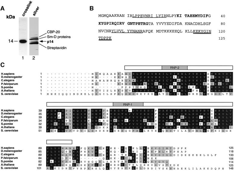
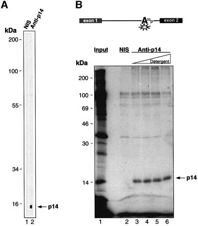
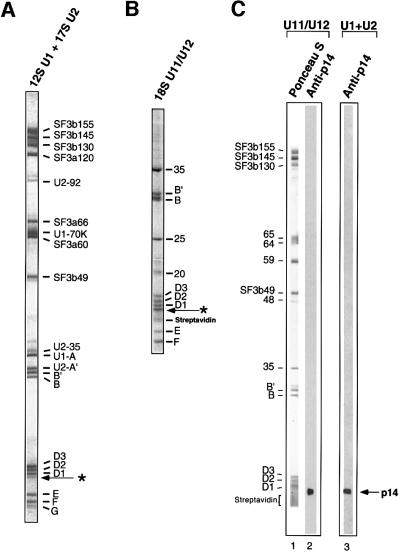
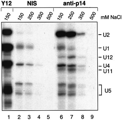
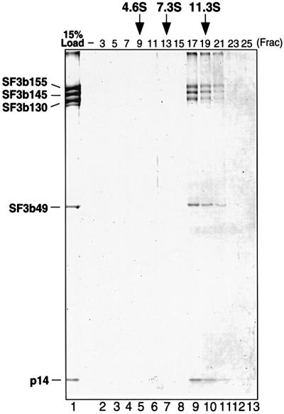
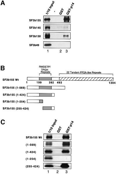
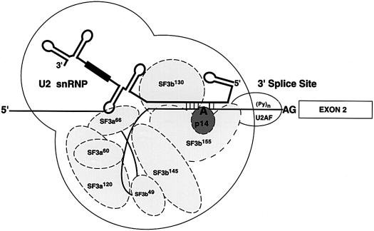
Previous UV cross-linking studies demonstrated that, upon integration of the U2 snRNP into the spliceosome, a 14 kDa protein (p14) interacts directly with the branch adenosine, the nucleophile for the first transesterification step of splicing. We have identified the cDNA encoding this protein by microsequencing a 14 kDa protein isolated from U2-type spliceosomes. This protein contains an RNA recognition motif and is highly conserved across species. Antibodies raised against this cDNA-encoded protein precipitated the 14 kDa protein cross-linked to the branch adenosine, confirming the identity of the p14 cDNA. A combination of immunoblotting, protein microsequencing and immunoprecipitation revealed that p14 is a component of both 17S U2 and 18S U11/U12 snRNPs, suggesting that it contributes to the interaction of these snRNPs with the branch sites of U2- and U12-type pre-mRNAs, respectively. p14 was also shown to be a subunit of the heteromeric splicing factor SF3b and to interact directly with SF3b155. Immuno precipitations indicated that p14 is present in U12-type spliceosomes, consistent with the idea that branch point selection is similar in the major and minor spliceosomes.


Figures

None
Fig. 1. p14 contains an RRM and is evolutionarily highly conserved. (A) The 14 kDa polypeptide region of purified U2-type spliceosomes. Proteins were separated on a 20% gel by SDS–PAGE and stained with silver (lane 2). For comparison, the branch site-cross-linked 14 kDa protein was separated in parallel and visualized by autoradiography (lane 1). (B) Amino acid sequence of human p14 (accession No. AF401310). Peptide sequences obtained by microsequencing of p14 from U2-dependent spliceosomes are indicated in bold, those from 17S U2 snRNPs are underlined, and those from 18S U11/U12 snRNPs are underlined twice. (C) Amino acid sequence alignment of human p14 and putative orthologs from D.melanogaster (#AC004767), C.elegans (#AF040642), P.falciparum (#AA550544/#AC004688), A.thaliana (#AB007727), S.pombe (#AL022299) and S.cerevisiae (#CAA86207.1). Residues identical in at least four sequences are boxed in black, and conserved residues (gray boxes) are grouped as follows: (D, E), (H, K, R), (A, F, I, L, M, P, V, W) and (C, G, N, Q, S, T, Y). The position of the RRM, including the highly conserved RNP-1 and RNP-2 motifs (shaded regions), is indicated above the alignment by an open bar. Sequence alignments were performed using the Clustal method and optimized by visual inspection.
None
Fig. 2. p14 is the 14 kDa protein cross-linked to the branch adenosine. (A) Specificity of anti-p14 antibodies. Nitrocellulose strips containing proteins isolated from nuclear extract were immunostained with pre-immune serum (NIS, lane 1) or anti-p14 antibodies (lane 2). (B) Immunoprecipitation (IP) verifies that the p14 cDNA encodes the 14 kDa branch site-cross-linked species. Benzophenone was placed on the branch adenosine, and a radioactive phosphate at the adjacent nucleotide, of a U2-type pre-mRNA (depicted schematically at the top). After allowing for splicing complex formation, cross-linking and subsequent IPs were performed. Immunoprecipitated cross-linked proteins were fractionated by SDS–PAGE on a 16% gel and visualized by autoradiography. Lane 1, total cross-links from nuclear extract; lane 2, IP using pre-immune serum; lanes 3–6, IPs using anti-p14 serum and increasing amounts of detergent prior to IP (lane 3, 0.05% NP-40; lane 4, 0.5% NP-40; lane 5, 0.5% NP-40 and 0.5% deoxycholate; lane 6, 0.5% NP-40, 0.5% deoxycholate and 0.1% SDS).
None
Fig. 3. 17S U2-enriched snRNPs and 18S U11/U12 snRNPs contain a low molecular weight protein recognized by p14-specific antibodies. (A) The protein composition of 17S U2 and 12S U1 snRNPs, or (B) 18S U11/U12 snRNPs (only low molecular weight proteins are shown). The putative p14 band is indicated by an arrow. Proteins were separated by SDS–PAGE on 10/13% gels and stained with Coomassie Blue. (C) Proteins from 18S U11/U12 snRNPs (lanes 1 and 2) or a mixture of 17S U2 and 12S U1 snRNPs (lane 3) were stained with Ponceau S (lane 1) or anti-p14 antibodies (lanes 2 and 3). The identities of the 18S U11/U12 proteins are indicated on the left.
None
Fig. 4. Antibodies directed against p14 immunoprecipitate both U2 snRNPs and U11/U12 snRNPs. Immunoprecipitations were performed in the presence of 150–500 mM NaCl (as indicated above each lane) with HeLa nuclear extract and pre-immune serum (lanes 2–5), anti-p14 serum (lanes 6–9) or the anti-Sm protein, monoclonal Y12 (lane 1). Immunoprecipitated snRNAs were 3′ end labeled with [32P]pCp, fractionated on a 7 M urea–10% polyacrylamide gel and visualized by autoradiography. The identity of the snRNA species was confirmed by northern blotting.
None
Fig. 5. p14 is a component of SF3b. SF3b was affinity purified with anti-SF3b155 antibodies and subjected to glycerol gradient centrifugation (see Materials and methods). Proteins were isolated from the affinity column eluate (lane 1, 15% of material loaded onto the gradient) or gradient fractions as indicated at the top of each lane (lanes 2–13) and analyzed as in Figure 3B. The identity of the bands, determined by western analysis, is indicated on the left. The S-values of human serum albumin (4.6), rabbit skeletal muscle aldolase (7.3) and bovine liver catalase (11.3), separated in parallel, are indicated at the top.
None
Fig. 6. p14 interacts directly with SF3b155 in GST pull-down assays. (A) GST pull-downs were performed with 35S-labeled, in vitro translated SF3b155, 145, 130 or 49 (as indicated on the left) and GST (lane 2) or GST–p14 (lane 3). Co-precipitated proteins or 10% of input (lane 1) were analyzed by SDS–PAGE on 10/13% gels and visualized by fluorography. (B) Schematic of SF3b155 domain structure and mutants used in (C). Regions containing RWDETP and TPGH repeats (shaded box) or tandem PP2A-like repeats (striped box) (Wang et al., 1998) are indicated. (C) Delineation of the p14 interaction domain in SF3b155. Wild-type SF3b155 or various deletion mutants (indicated on the left) were incubated with GST (lane 2) or GST–p14 (lane 3), and glutathione–Sepharose-precipitated proteins or 10% of input were analyzed as in (A).
None
Fig. 7. p14 is present in both major and minor spliceosomes. (A) Anti-p14 antibodies precipitate U2-dependent spliceosomes. Splicing was performed for 5 (lanes 1–3) or 20 min (lanes 4–6) with MINX pre-mRNA and immunoprecipitations (IPs) carried out with pre-immune (NIS; lanes 2 and 5) or anti-p14 (lanes 3 and 6) serum. Twenty percent of the splicing reaction used for IP is also shown (lanes 1 and 4). (B) Anti-p14 antibodies precipitate U12-dependent spliceosomes. Splicing was performed for 30 (lanes 1–6) or 240 min (lanes 7–12) with P120 pre-mRNA in the presence of an oligonucleotide against U2 (lanes 1–3 and 7–9) or U12 (lanes 4–6 and 10–12). IPs were carried out with pre-immune (NIS; lanes 2, 5, 8 and 11) or anti-p14 (lanes 3, 6, 9 and 12) serum. Twenty percent of the splicing reaction used for IP is also shown (lanes 1, 4, 7 and 10). RNA was analyzed as described in Materials and methods and visualized by autoradiography.
None
Fig. 8. Model of p14 interactions at the branch site in pre-spliceosomes. The U2 snRNP including U2 snRNA (thick solid line) and its Sm site (black box), and a subset of U2-specific proteins (ellipses) are shown schematically. Exon 2 is depicted by a box, intron sequences by a thin solid line, the polypyrimidine tract by (Py)n and the branch adenosine (A) and 3′ splice site AG dinucleotide are in bold.

References

    1. Abovich N. and Rosbash,M. (1997) Cross-intron bridging interactions in the yeast commitment complex are conserved in mammals. Cell, 89, 403–412. - PubMed
    1. Behrens S.E., Tyc,K., Kastner,B., Reichelt,J. and Lührmann,R. (1993) Small nuclear ribonucleoprotein (RNP) U2 contains numerous additional proteins and has a bipartite RNP structure under splicing conditions. Mol. Cell. Biol., 13, 307–319. - PMC - PubMed
    1. Bennett M., Michaud,S., Kingston,J. and Reed,R. (1992) Protein components specifically associated with prespliceosome and spliceosome complexes. Genes Dev., 6, 1986–2000. - PubMed
    1. Berglund J.A., Chua,K., Abovich,N., Reed,R. and Rosbash,M. (1997) The splicing factor BBP interacts specifically with the pre-mRNA branchpoint sequence UACUAAC. Cell, 89, 781–787. - PubMed
    1. Brosi R., Hauri,H.P. and Kramer,A. (1993) Separation of splicing factor SF3 into two components and purification of SF3a activity. J. Biol. Chem., 268, 17640–17646. - PubMed

Publication types

MeSH terms

Associated data