. 2001 Aug 15;20(16):4603-17.
doi: 10.1093/emboj/20.16.4603.

CENP-H, a constitutive centromere component, is required for centromere targeting of CENP-C in vertebrate cells

Affiliations

CENP-H, a constitutive centromere component, is required for centromere targeting of CENP-C in vertebrate cells

T Fukagawa et al. EMBO J. .

Abstract

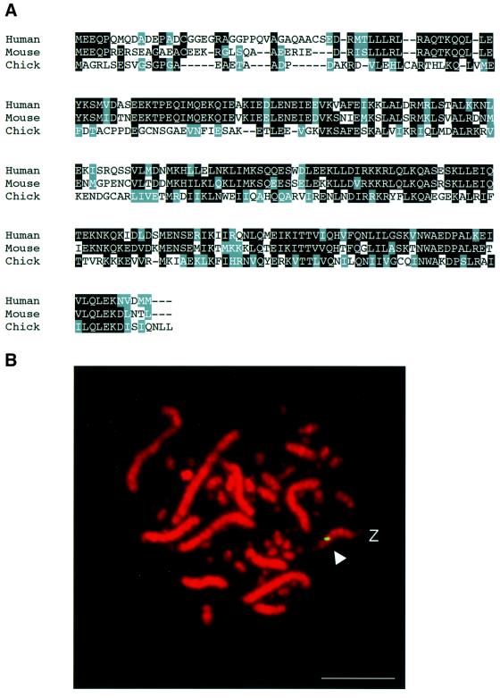
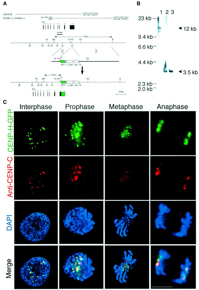
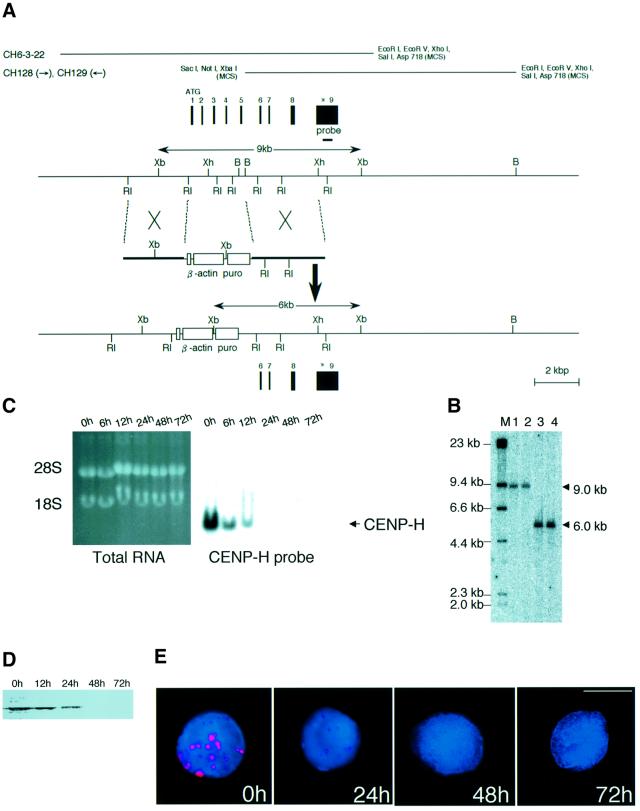
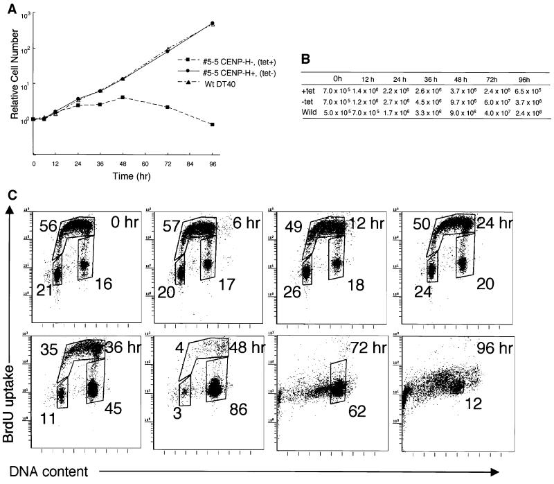
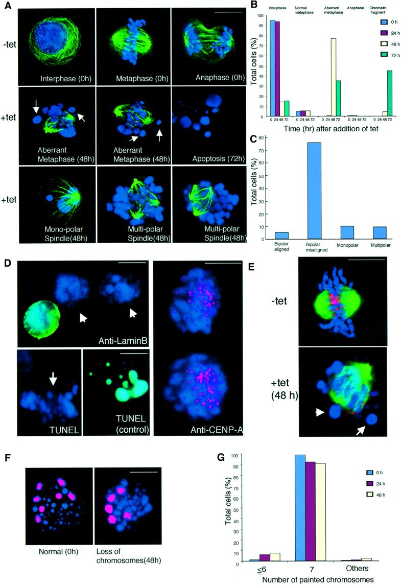
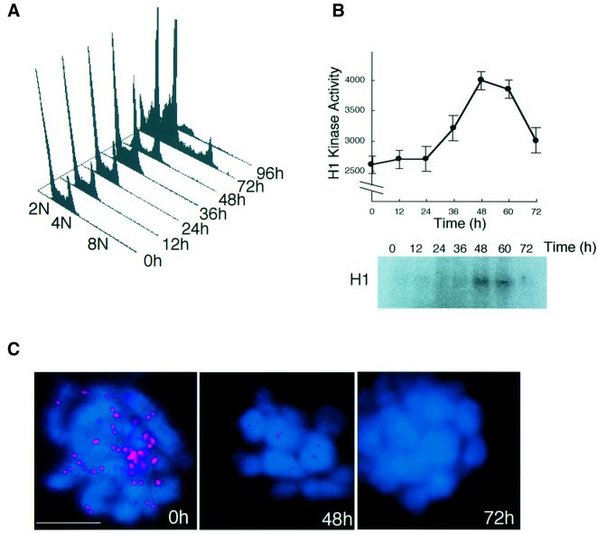
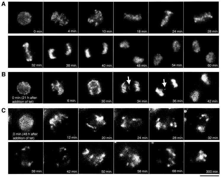
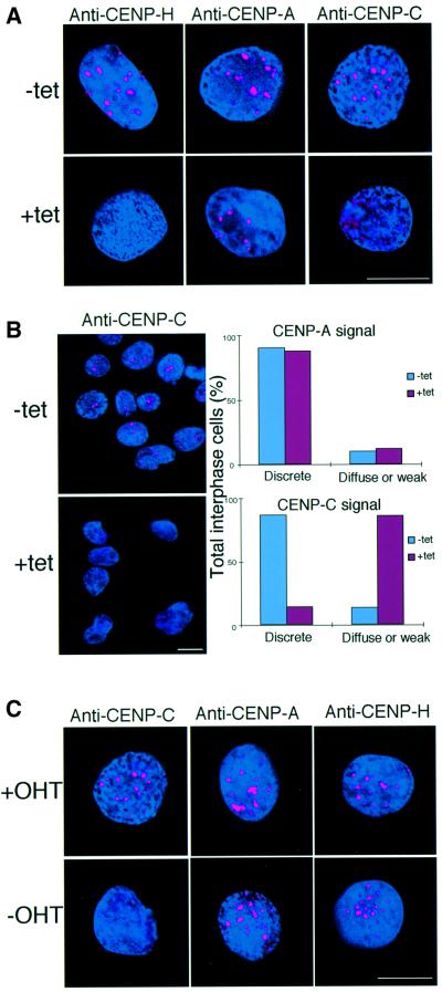
CENP-H has recently been discovered as a constitutive component of the centromere that co-localizes with CENP-A and CENP-C throughout the cell cycle. The precise function, however, remains poorly understood. We examined the role of CENP-H in centromere function and assembly by generating a conditional loss-of-function mutant in the chicken DT40 cell line. In the absence of CENP-H, cell cycle arrest at metaphase, consistent with loss of centromere function, was observed. Immunocytochemical analysis of the CENP-H-deficient cells demonstrated that CENP-H is necessary for CENP-C, but not CENP-A, localization to the centromere. These findings indicate that centromere assembly in vertebrate cells proceeds in a hierarchical manner in which localization of the centromere-specific histone CENP-A is an early event that occurs independently of CENP-C and CENP-H.


Figures

None
Fig. 1. Identification of the chicken CENP-H gene. (A) Comparison of amino acid sequences of human, mouse and chicken CENP-H proteins. Amino acid sequence is shown as a single-letter code. Amino acids that are conserved between species are shaded. Nucleotide sequence of the chicken CENP-H cDNA is available from DDBJ/EMBL/GenBank (accession No. AB055783). (B) Chromosomal localization of the CENP-H gene by FISH. DT40 metaphase spreads were hybridized with the CENP-H probe. The arrowhead indicates a probe-specific hybridization signal (green). DNA is stained with 4′,6-diamidine-2-phenylindole (DAPI) and colored red. CENP-H was mapped to chromosome Z, indicating that there is a single CENP-H allele in each DT40 cell. The scale bar corresponds to 10 µm.
None
Fig. 2. Chicken CENP-H localizes to centromeres throughout the cell cycle. (A) Restriction maps of the CENP-H gene, the CENP-H–GFP replacement construct and the configuration of the targeted locus. Black boxes indicate the positions of exons. Exon 9, which contains the stop codon, was replaced with a portion of the sequence of exon 9 fused to a GFP tag followed by the neo-resistance gene (neo) and 3′ homology region. Relevant restriction enzyme sites shown are as follows: RI, EcoRI; Xh, XhoI; B, BamHI; RV, EcoRV. The position of the probe used for Southern hybridiza tion is indicated. A new 3.5 kb BamHI fragment hybridizes with the probe if targeted integration of the indicated replacement construct occurs. (B) Restriction analysis of the CENP-H–GFP allele. Genomic DNAs from wild-type DT40 cells (lane 1) and the targeted clones (lanes 2 and 3) were digested with BamHI, size fractionated by 0.7% agarose gel electrophoresis, transferred to a nylon filter and hybridized with the probe indicated in (A). (C) Localization of CENP-H–GFP at progressive stages of the cell cycle in DT40 cells. Cells were fixed and stained with anti-CENP-C antibody (red), and green signals are specific for CENP-H–GFP. Nuclei and chromosomes were visualized by counterstaining with DAPI (blue). The scale bar corresponds to 10 µm. As shown in the merged images, CENP-H–GFP signals are co-localized with CENP-C signals throughout the cell cycle.
None
Fig. 3. Generation of a ΔCENP-H clone carrying a chicken CENP-H transgene under the control of a tet-repressible promoter. (A) Restriction maps of the CENP-H locus, the gene disruption construct and configuration of the targeted locus. The targeted construct is expected to disrupt exons 1–5. Relevant restriction enzyme sites shown are as follows: RI, EcoRI; Xh, XhoI; B, BamHI; Xb, XbaI. The position of the probe used for Southern hybridization is indicated. A new 6.0 kb XbaI fragment hybridizes with the probe if targeted integration of the construct occurs. (B) Restriction analysis of the targeted integration of the CENP-H disruption construct. Genomic DNAs from wild-type DT40 cells (CENP-H+, lane 1), a clone with random integration of the CENP-H transgene (CENP-H+/CENP-H transgene, lane 2) and targeted clones (CENP-H–/CENP-H transgene, lanes 3 and 4) were digested with XbaI, size fractionated by 0.7% agarose gel electrophoresis, transferred to a nylon filter and hybridized with the probe indicated in (A). (C) Suppression of CENP-H expression from the transgene. Total RNA was isolated from #5-5 cells at the times indicated following addition of tet. RNA samples (20 µg/lane) were fractionated on formaldehyde gels (left side of figure). Northern blot hybridization was carried out with the full-length CENP-H cDNA as the probe. Bands other than that for CENP-H band were not detected, indicating that the truncated form of CENP-H was not expressed. (D) Western blot analysis with anti-CENP-H antibody of the #5-5 cell extract at the times indicated following addition of tet. (E) Immunofluorescence analysis of #5-5 cells at times indicated following the addition of tet using anti-CENP-H antibody (red). DNA was counterstained with DAPI (blue). The scale bar corresponds to 10 µm.
None
Fig. 4. CENP-H is essential for normal progression of the cell cycle. (A) Representative growth curves for the cell cultures indicated. Tet was added at time 0 to the #5-5 CENP-H (tet+) culture. The number of cells not stained with Trypan blue was counted. Each experiment was performed twice, and each time point was examined in duplicate. (B) Total cell number plotted in (A). (C) Cell cycle distribution of #5-5 cells following inhibition of CENP-H transgene expression by adding tet at time 0. Cells were stained with FITC–anti-BrdU (y-axis, log scale) to detect BrdU incorporation and with propidum iodide to detect total DNA (x-axis, linear scale). The lower-left box represents G1-phase cells, the upper box represents S-phase cells, and the lower-right box represents G2–M-phase cells. The numbers given in the boxes indicate the percentage of gated events.
None
Fig. 5. CENP-H-deficient cells show metaphase arrest associated with aberrant chromosomes and spindles that lead to chromosome missegregation. (A) Chromosome morphology and α-tubulin staining (green) in the absence or presence of tet. DNA was counterstained with DAPI (blue). In the absence of tet, cells show the normal staining pattern for α-tubulin (upper three panels). In the presence of tet, cells in which chromosomes were not aligned at the metaphase plate were observed. Arrows indicate the misaligned chromosomes (middle panels) 48 h after addition of tet. Cells with apoptosis were observed 72 h after addition of tet (middle right panel). We also detected cells with monopolar and multipolar spindles (lower panels). The scale bar corresponds to 10 µm. (B) Quantitation of aberrant #5-5 cells following inhibition of CENP-H transgene expression after addition of tet at time 0. We scored the number of interphase cells, normal metaphase cells, aberrant metaphase cells described in (A), anaphase cells and cells with micronuclei. We scored ∼2000 cells at each time point. (C) Quantitation of cells with misaligned chromosomes 48 h after addition of tet. Misaligned chromosomes were scored as bipolar spindle with misaligned chromosomes, monopolar spindle or multispindle. (D) Immunofluorescence analysis of #5-5 cells 48 h after addition of tet with anti-LaminB antibody (upper left). Aberrant chromosomes (arrow) are not stained. Apoptotic cells can be detected by TUNEL assay (control). Aberrant chromosomes (arrow) were not stained (lower left). Immunofluorescence analysis of #5-5 cells 72 h after addition of tet with anti-CENP-A antibody (right). Aberrant chromosomes are stained with CENP-A antibody (red). (E) Double staining #5-5 cells 0 (–tet) or 48 h (+tet) after addition of tet with anti-α-tubulin (green) and anti-CENP-A (red) antibodies. Arrows indicate misaligned chromo somes that do not appear to attach to microtubules, although many other chromosomes appear to have formed microtubule attachments. (F) To examine chromosome loss, we used FISH analysis with chromosome-specific painting probes. We used painting probes for chicken chromosomes 1, 2 and 3. Because DT40 cells have three copies of chromosome 2, we detected seven painted chromosomes in normal cells (left panel). #5-5 cells with loss of chromosomes (right panel) were detected after addition of tet. The scale bars correspond to 10 µm. (G) Distribution of the number of painted chromosomes per cell. #5-5 cells were cultured after addition of tet. At the indicated time, cells were treated with colcemid for 3 h. The number of painted chromosomes was scored in ∼200 metaphase cells.
None
Fig. 6. CENP-H-deficient cells proceed through the cell cycle without normal chromosome segregation. (A) FACS analysis of the cell cycle pattern of #5-5 cells after addition of tet. The positions of cell populations with 2N, 4N or 8N DNA content are illustrated. At 72 h, a peak of 8N DNA content is observed. (B) Histone H1 kinase activity in chicken p34CDC2 immunoprecipitates was measured in the cells isolated in (A). Kinase activity was measured using purified histone H1 as a substrate. Total radioactivity of histone H1 was measured with a STORM imager. (C) Immunofluorescence analysis of metaphase #5-5 cells 0, 48 or 72 h after addition of tet with antibody against an outer centromere protein ZW10 (red). At 0 h (control), cells were treated with colcemid for 3 h. DNA was counterstained with DAPI (blue). The scale bar corresponds to 10 µm.
None
Fig. 7. Dynamics of chromosomes by time-lapse observation. (A) Selected frames of chromosomes from prophase to telophase in #5-5 cells (–tet). The numbers at the bottom of each image represent the time in minutes. The scale bar corresponds to 10 µm. (B) From 21 h after addition of tet, one prophase cell was followed for time-lapse observations at intervals of 2 min. Selected frames are shown. A lagging chromosome can be observed (arrow). (C) From 48 h after addition of tet, one prophase cell was followed for time-lapse observation. Once this cell entered mitosis, the cell was arrested at metaphase for >300 min.
None
Fig. 8. CENP-H is required for centromere targeting of CENP-C but not CENP-A. (A) Immunofluorescence analysis of #5-5 cells 0 (–tet) or 48 h (+tet) after addition of tet. Interphase cells were stained with anti-CENP-H, anti-CENP-A and anti-CENP-C antibodies. Antibody signals were detected with Cy3-conjugated secondary antibodies (red). DNA was counterstained with DAPI (blue). The scale bars correspond to 10 µm. (B) Quantitation of interphase cells with diffuse signals of CENP-A and CENP-C. The left panel provides an example of the diffuse CENP-C signals in CENP-H-deficient cells. As shown in the left panel, many #5-5 interphase cells displayed diffuse CENP-C signals at 48 h after addition of tet, while discrete CENP-C signals were observed in the absence of tet. The right panel shows quantitative data. We scored ∼200 interphase cells. (C) Immunofluorescence analysis of Δ/CENP-C-ER cells grown in the presence (+OHT) or absence (–OHT, restrictive conditions) of OHT for 72 h. Rabbit antibodies to anti-CENP-C, anti-CENP-A or anti-CENP-H were applied to interphase cells and detected with Cy3-conjugated second antibodies (red). DNA was counterstained with DAPI (blue).

References

    1. Amon A. (1999) The spindle checkpoint. Curr. Opin. Genet. Dev., 9, 69–75. - PubMed
    1. Brown M.T. (1995) Sequence similarities between the yeast chromosome segregation protein Mif2 and the mammalian centromere protein CENP-C. Gene, 160, 111–116. - PubMed
    1. Brown M.T., Goetsch,L. and Hartwell,L.H. (1993) MIF2 is required for mitotic integrity during anaphase spindle elongation in Saccharomyces cerevisiae. J. Cell Biol., 123, 387–403. - PMC - PubMed
    1. Buerstedde J.M. and Takeda,S. (1991) Increased ratio of targeted to random integration after transfection of chicken B cell lines. Cell, 67, 179–188. - PubMed
    1. Chan G.K., Jablonski,S.A., Starr,D.A., Goldberg,M.L. and Yen,T.J. (2000) Human Zw10 and ROD are mitotic checkpoint proteins that bind to kinetochores. Nature Cell Biol., 2, 944–947. - PubMed

Publication types

MeSH terms