. 2001 Sep;69(9):5318-28.
doi: 10.1128/IAI.69.9.5318-5328.2001.

Independent and coordinate effects of ADP-ribosyltransferase and GTPase-activating activities of exoenzyme S on HT-29 epithelial cell function

Affiliations

Independent and coordinate effects of ADP-ribosyltransferase and GTPase-activating activities of exoenzyme S on HT-29 epithelial cell function

J E Fraylick et al. Infect Immun. 2001 Sep.

Abstract

Type III-mediated translocation of exoenzyme S (ExoS) into HT-29 epithelial cells by Pseudomonas aeruginosa causes complex alterations in cell function, including inhibition of DNA synthesis, altered cytoskeletal structure, loss of readherence, microvillus effacement, and interruption of signal transduction. ExoS is a bifunctional protein having both GTPase-activating (GAP) and ADP-ribosyltransferase (ADPRT) functional domains. Comparisons of alterations in HT-29 cell function caused by P. aeruginosa strains that translocate ExoS having GAP or ADPRT mutations allowed the independent and coordinate functions of the two activities to be assessed. An E381A ADPRT mutation revealed that ExoS ADPRT activity was required for effects of ExoS on DNA synthesis and long-term cell rounding. Conversely, the R146A GAP mutation appeared to have little impact on the cellular effects of ExoS. While transient cell rounding was detected following exposure to the E381A mutant, this rounding was eliminated by an E379A-E381A ADPRT double mutation, implying that residual ADPRT activity, rather than GAP activity, was effecting transient cell rounding by the E381A mutant. To explore this possibility, E381A and R146A-E381A mutants were examined for their ability to ADP-ribosylate Ras in vitro or in vivo. While no ADP-ribosylation of Ras was detected by either mutant in vitro, both mutants were able to modify Ras when translocated by the bacteria, with the R146A-E381A mutant causing more efficient modification than the E381A mutant, in association with increased inhibition of DNA synthesis. Comparisons of Ras ADP-ribosylation by wild-type and E381A mutant ExoS by two-dimensional electrophoresis found the former to ADP-ribosylate Ras at two sites, while the latter modified Ras only once. These studies draw attention to the key role of ExoS ADPRT activity in causing the effects of bacterially translocated ExoS on DNA synthesis and cell rounding. In addition, the studies provide insight into the enhancement of ExoS ADPRT activity within the eukaryotic cell microenvironment and into possible modulatory roles that the GAP and ADPRT domains might have on the function of each other.


Figures

FIG. 1
FIG. 1
ExoS cross-reactive protein produced by plasmid constructs. An ExoS immunoblot of culture supernatants from strains expressing the indicated construct is shown. (See also Tables 1 and 2).
FIG. 2
FIG. 2
Inhibition of DNA synthesis associated with ExoS ADP-ribosyltransferase activity. HT-29 cells were seeded at 105 cells/ml and grown for 48 h to ∼40% confluency. Cell culture medium was then removed and replaced with McCoys plus 0.6% BSA medium containing no bacteria (“0”) or 107 CFU of strain 388 (388), non-ExoS-producing strain 388ΔS (388ΔS), or of PA103ΔUT strains containing the pUCP control (pUCP), the pUCP-ExoS E381A ADPRT mutant (E381A), or the pUCP-ExoS (ExoS) vectors. Following a 4-h coculture period, bacteria were removed and replaced with medium containing [3H]thymidine and antibiotics to inhibit further bacterial growth and then assayed for DNA synthesis after 20 h. The results are expressed as the percent [3H]thymidine incorporation relative to nonbacterially treated control cells, and the means and standard errors of the assays performed in triplicate from three independent studies are shown.
FIG. 3
FIG. 3
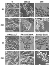
Effects of ExoS ADP-ribosyltransferase activity on transient and long-term alterations in cell morphology. HT-29 cells were grown and cocultured as described in Fig. 2 with no bacteria (“0”) strains 388 and 388ΔS, or PA103ΔUT strains containing the indicated vector. Cells were examined for alterations in morphology by phase-contrast microscopy following removal of bacteria after a 4-h coculture period (4h). Long-term effects on morphology were assessed by culturing cells for an additional 20 h after removal of bacteria in medium containing antibiotics (24h). Transient cell rounding was detected following coculture with strains 388ΔS and PA103ΔUT-E381A, while severe cell rounding persisted for 24 h in cells cocultured with ADPRT-active ExoS produced by strains 388 and PA103ΔUT-ExoS. The images shown are representative of three or more independent studies.
FIG. 4
FIG. 4
Inhibition of HT-29 cell DNA synthesis caused by ExoS GAP and ADPRT mutants. HT-29 cells were seeded and cocultured with the indicated bacterial strains for 4 h and assayed for DNA synthesis as described in Fig. 2. Strains examined in parallel in these studies include PA103ΔUT containing the pUCP vector control (pUCP), pUCP-ExoS (ExoS), pUCP-ExoS with an R146A GAP mutation (146), pUCP-ExoS with an E381A ADPRT mutation (381), pUCP-ExoS with both an R146A GAP and an E381A ADPRT mutation (146/381), pUCP-ExoS with an E379A-E381A ADPRT double mutation (379/381), and pUCP-ExoS with an R146A GAP and E379A-E381A double ADPRT mutation (146/379/381). Results are expressed as the percentage of [3H]thymidine incorporation relative to nonbacterially treated control cells, and the means and standard deviations of the assays performed in triplicate from a representative experiment of three independent studies are shown.
FIG. 5
FIG. 5
Effects of ExoS GAP and ADPRT mutations on transient and long-term alterations in cell morphology. HT-29 cells were grown and cocultured with no bacteria (“0”) or strain PA103ΔUT containing the indicated ExoS construct as described in Fig. 4 and assayed for transient (4h) or long-term (24h) effects on cell morphology, as described in Fig. 3. In (A) Transient cell rounding was detected following coculture with strains containing ExoS, E381A, and R146A, with less rounding caused by the R146A-E381A GAP-ADPRT mutant. Cell rounding persisted at 24 h in cells cocultured with ADPRT-active ExoS and the R146A mutant. (B) Comparisons of morphological alterations caused by the E379A-E381A double ADPRT and R146A-E379A-E381A GAP-double ADPRT mutants found minimal transient or long-term cell rounding. The images shown are from analyses performed in parallel and are representative of multiple independent studies.
FIG. 6
FIG. 6
ADP-ribosylation of Ras by ExoS GAP and ADPRT mutants in vitro and in vivo. (A) In vitro. ADP-ribosylation of purified recombinant H-Ras was examined in reaction mixtures containing 0.5 μM H-Ras, 10 mM NAD, 0.2 μM 14-3-3ξ, and 30 μl of culture supernatant from PA103ΔUT strains producing the indicated ExoS construct. Reactions were allowed to proceed for 1 h at room temperature, stopped with 4× Laemmli buffer and heating at 95°C, resolved by SDS–12% PAGE, blotted onto PVDF membrane, probed with mouse anti-H-Ras LA069 and anti-pan Ras LA045 antibodies, and visualized by ECL. A reaction containing purified ExoS (S) was included as a positive control. (B) In vivo. ADP-ribosylation of cellular Ras was examined following a 5-h coculture of HT-29 cells with PA103ΔUT containing the indicated ExoS plasmid construct. Following removal of bacteria, cells were lysed in TBS-TDS (10 mM Tris [pH 7.4], 140 mM NaCl, 1% Triton X-100, 0.5% sodium deoxycholate, 0.1% SDS), and Ras was immunoprecipitated from cell extracts using Y13-259 Ras antibody and protein G-agarose. Immunoprecipitates were resolved by SDS–12% PAGE, transferred to PVDF membranes, and Ras was detected and visualized as in panel A. The mobility of modified (m) and unmodified (u) Ras is indicated. The two bands observed for unmodified Ras in vivo represent the different Ras species, while the two bands observed for modified Ras in vivo reflect differences in the efficiency of Ras modification.
FIG. 7
FIG. 7
2DE analysis of the in vivo ADP-ribosylation of Ras by ExoS GAP and ADPRT mutants. Following a 5-h coculture of HT-29 cells with strain PA103ΔUT producing the indicated ExoS protein, Ras was immunoprecipitated from cell extracts, as in Fig. 6B. Ras immunoprecipitates were solubilized in 2DE rehydration buffer, as described in Materials and Methods, and focused on Immobiline pH 4 to 7 gradient gel strips. Strips were equilibrated in SDS equilibration buffer, proteins were resolved in the second dimension on SDS–12% polyacrylamide gels and transferred to PVDF membranes, and Ras was detected as in Fig. 6. The mobility of unmodified (U) and modified (M) Ras is indicated. Multiple spots represent the different Ras isoforms. pI and molecular mass markers (in kilodaltons [kD], are indicated above and to the left of each panel, respectively.
FIG. 8
FIG. 8
Monitoring the translocation ExoS mutant protein and ADPRT activity in HT-29 cells. HT-29 cells, plated in 100-mm dishes, as described in Fig. 2, were cocultured with PA103ΔUT-ExoS mutant strains for 4.5 h, permeabilized with 0.2% saponin, and then fractionated into cytosolic and membrane components using a modification of the method of Kenny and Finlay (24), as detailed in Materials and Methods. The three fractions obtained include (i) the cytosolic fraction (C), which contains cytosolic, microsomal, and plasma membrane proteins; (ii) the TX-100 fraction (TX), which contains membrane-solubilized proteins; and (iii) the insoluble pellet fraction (P), which includes adherent bacteria and host cell cytoskeleton and nuclei. (A) Analysis of ExoS in bacterial culture supernatants. A 10-μl volume of TSBD-N culture supernatants of the indicated bacterial strain was resolved on SDS–15% polyacrylamide gels and immunoblotted for ExoS or assayed for ExoS ADPRT activity, as in Table 1, prior to the processing of bacteria and coculture with HT-29 cells. An ExoS-ExoT standard (Std), quantified to have 50 ng of ExoS protein, was included in the immunoblot. (B) Analysis of ExoS in HT-29 cell fractions. A 50-μl volume of cytosolic (C), TX-100 (TX), or pellet (P) fractions of HT-29 cells, following coculture with the indicated bacterial strains, was resolved on SDS-PAGE and immunoblotted for ExoS. A 10-μl volume of the respective fraction was assayed for ExoS ADPRT in parallel with culture supernatants assayed in panel A. A 20-μl volume of PA103-ExoS culture supernatant (S) was included in immunoblot analyses as a positive control. (C) Analysis of Ral modification in HT-29 cell fractions. To directly compare ExoS protein levels to functional substrate modification, ExoS blots were reprobed for Ral, using a monoclonal anti-RalA antibody (Transduction Laboratories), followed by HRP-conjugated anti-mouse IgG, and then visualized by ECL. Asterisks identify modified Ral bands. The mean ± the standard deviation of the ExoS ADPRT activity of the respective samples is indicated below the immunoblots in panels A and B and is expressed as femtomoles of ADP-ribose transferred per minute per microliter of sample. ExoS, ExoT, and the mobility of modified (m) and unmodified (u) Ral are labeled, as are the molecular mass references.

Similar articles

Cited by

References

    1. Albright C F, Giddings B W, Liu J, Vito M, Wienberg R A. Characterization of a guanine nucleotide dissociation stimulator for a ras-related GTPase. EMBO J. 1993;12:339–347. - PMC - PubMed
    1. Chuang S E, Chen A L, Chao C C. Growth of Escherichia coli and how temperature dramatically increases the transformation frequency by electroporation. Nucleic Acid Res. 1995;23:1641. - PMC - PubMed
    1. Coburn J, Dillon S T, Iglewski B H, Gill D M. Exoenzyme S of Pseudomonas aeruginosa ADP-ribosylates the intermediate filament protein vimentin. Infect Immun. 1989;57:996–998. - PMC - PubMed
    1. Coburn J, Gill D M. ADP-ribosylation of p21ras and related proteins by Pseudomonas aeruginosa exoenzyme S. Infect Immun. 1991;59:4259–4262. - PMC - PubMed
    1. Coburn J, Wyatt R T, Iglewski B H, Gill D M. Several GTP-binding proteins, including p21c-H-ras, are preferred substrates of Pseudomonas aeruginosa exoenzyme S. J Biol Chem. 1989;264:9004–9008. - PubMed

Publication types

LinkOut - more resources