. 2001 Sep;69(9):5671-8.
doi: 10.1128/IAI.69.9.5671-5678.2001.

Expression of a Mycobacterium tuberculosis arabinomannan antigen in vitro and in vivo

Affiliations

Expression of a Mycobacterium tuberculosis arabinomannan antigen in vitro and in vivo

J R Schwebach et al. Infect Immun. 2001 Sep.

Abstract

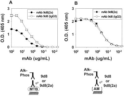
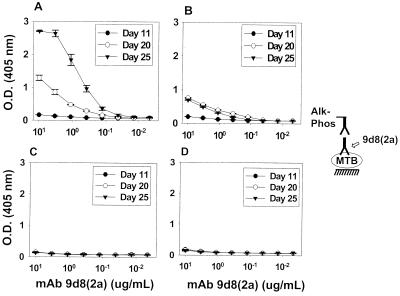
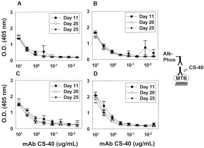
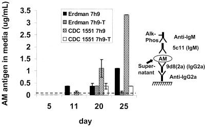
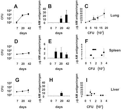
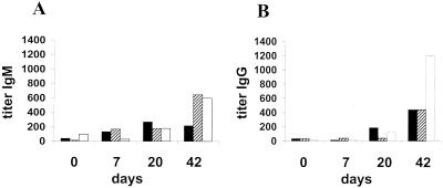
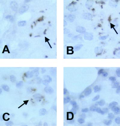
The outermost layer of Mycobacterium tuberculosis contains two major polysaccharides, arabinomannan (AM) and glucan (GC). We studied the in vitro and in vivo expression of an M. tuberculosis AM antigen using monoclonal antibody (MAb) 9d8 (2a), an isotype-switched variant of the immunoglobulin G3 (IgG3) MAb 9d8. MAb 9d8 had been previously shown to bind M. tuberculosis AM and the M. tuberculosis surface. Our in vitro experiments showed that MAb 9d8(2a) bound strongly to whole-cell M. tuberculosis Erdman but not to the CDC 1551 strain grown in medium for an extended period. However, AM antigen was detected in the culture supernatant of both strains, and its concentration increased in a time-dependent manner. The detection of AM antigen from both strains was decreased in the presence of Tween 80. In mice infected with M. tuberculosis Erdman, AM antigen accumulated in organ homogenates concomitant to an increase in bacterial organ burden and an increase in IgG and IgM titer to AM. These results (i) indicate that the surface expression of AM during in vitro growth changes with culture age, is strain dependent, and is affected by the presence of Tween 80 in the culture media; (ii) show that AM is produced by bacteria growth in vivo; and (iii) demonstrate that the amount of in vivo-detected AM can be dependent on the number of bacteria in the infected organ.


Figures

FIG. 1
FIG. 1
ELISA of MAbs 9d8(2a) and 9d8 binding to M. tuberculosis and AM. MAb 9d8(2a) or 9d8 was serially diluted in an ELISA using M. tuberculosis Erdman (A) or purified AM (B). Each value represents the average of three measurements. Error bars show the standard deviations of the means. These growth experiments and the ELISA were repeated twice with similar results. O.D., optical density; MTB, M. tuberculosis; Alk-Phos, alkaline phosphatase.
FIG. 2
FIG. 2
Binding of MAb 9d8(2a) to whole-cell M. tuberculosis Erdman and CDC 1551 at various culture times. M. tuberculosis Erdman (A and B) and CDC 1551 (C and D) were grown in the absence (A and C) or the presence (B and D) of Tween for periods of 11, 20, or 25 days before harvest. Each value represents the average of three measurements. Error bars show the standard deviations of the means. These growth experiments and the ELISA were repeated twice with similar results. O.D., optical density; MTB, M. tuberculosis; Alk-Phos; alkaline phosphatase.
FIG. 3
FIG. 3
Binding of MAb CS-40 to whole-cell M. tuberculosis Erdman and CDC 1551 at various culture times. M. tuberculosis Erdman (A and B) and CDC 1551 (C and D) were grown in the absence (A and C) or the presence (B and D) of Tween for periods of 11, 20, or 25 days before harvest. Each value represents the average of three measurements. Error bars show the standard deviations of the means. These growth experiments and the ELISA were repeated twice with similar results. O.D., optical density; MTB, M. tuberculosis; Alk-Phos; alkaline phosphatase.
FIG. 4
FIG. 4
Capture ELISA of AM in M. tuberculosis culture medium. M. tuberculosis Erdman and CDC 1551 were grown in the absence or presence of Tween 80 for periods of 5, 11, 15, 20, or 25 days before harvest of bacterial supernatant. Each value represents the average of two separate measurements. Error bars represent the standard deviation of the means. These growth experiments and the ELISA were repeated twice with similar results. The detection limit of the assay is indicated by the dotted line. Alk-Phos, alkaline phosphatase.
FIG. 5
FIG. 5
Capture ELISA of AM in organ homogenate compared to organ CFU. The number of CFU in the lungs (A), spleens (D), or livers (G) of mice infected with M. tuberculosis Erdman was determined 7, 20, and 42 days after infection. Each value represents the average of three animals. Error bars show the standard deviation of the mean. Organ homogenates were assayed for micrograms of MAb 9d8(2a) AM antigen/organ in the lung (B), spleen (E), or liver (H) 0, 7, 20, and 42 days after infection. The detection limit of the assay is indicated by the dotted line. Each value represents the average of the three animals, using two measurements for each animal. Error bars represent the standard deviation of all six measurements. The CFU data for each animal organ were also related to the number of micrograms of MAb 9d8(2a) AM antigen in that animal organ (C, F, and I) for all day-7 (⧫), day-20 (●), and day-42 (■) mice. Each value represents the average of the two measurements of MAb 9d8(2a) AM antigen concentration per animal organ, and error bars represent the standard deviation of those two measurements. The point (⧫) at the bottom left of Panel C represents three mice that have the same apparent graphic value.
FIG. 6
FIG. 6
Antibody titer to AM during M. tuberculosis infection. Murine sera were assayed for IgM (A) and IgG (B) antibody titer to AM 0, 7, 20, and 42 days after infection of mice with M. tuberculosis Erdman. The three animals assayed for antibody titer at each period were the same animals used to generate the data for Fig. 5.
FIG. 7
FIG. 7
MAb 9d8 immunohistochemistry. Infected (day 42) (A to C) and uninfected (day 0) (D) murine lung samples were stained for the AM antigen using MAb 9d8 and horseradish peroxidase-conjugated secondary antibody. Arrows indicate bacterial shapes (A and B) or more diffuse staining (C).

Similar articles

Cited by

References

    1. Casadevall A, Mukherjee J, Scharff M D. Monoclonal antibody based ELISAs for cryptococcal polysaccharide. J Immunol Methods. 1992;154:27–35. - PubMed
    1. Chatterjee D, Khoo K H. Mycobacterial lipoarabinomannan: an extraordinary lipoheteroglycan with profound physiological effects. Glycobiology. 1998;8:113–120. - PubMed
    1. Chatterjee D, Lowell K, Rivoire B, McNeil M R, Brennan P J. Lipoarabinomannan of Mycobacterium tuberculosis. Capping with mannosyl residues in some strains. J Biol Chem. 1992;267:6234–6239. - PubMed
    1. Coates S R, Hansen D, Schecter G, Slutkin G, Hopewell P, Affronti L, Echenberg D F. Identification of Mycobacterium tuberculosis antigens in Seibert fractions by immunoblotting. J Clin Microbiol. 1986;24:126–130. - PMC - PubMed
    1. Cooper L J, Shikhman A R, Glass D D, Kangisser D, Cunningham M W, Greenspan N S. Role of heavy chain constant domains in antibody-antigen interaction. Apparent specificity differences among streptococcal IgG antibodies expressing identical variable domains. J Immunol. 1993;150:2231–2242. - PubMed

Publication types

MeSH terms

LinkOut - more resources