

Cholera toxin B subunit as a carrier molecule promotes antigen presentation and increases CD40 and CD86 expression on antigen-presenting cells
- PMID: 11500448
- PMCID: PMC98688
- DOI: 10.1128/IAI.69.9.5716-5725.2001
Cholera toxin B subunit as a carrier molecule promotes antigen presentation and increases CD40 and CD86 expression on antigen-presenting cells
Abstract
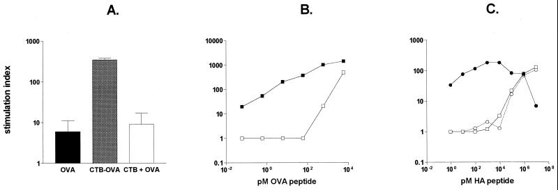
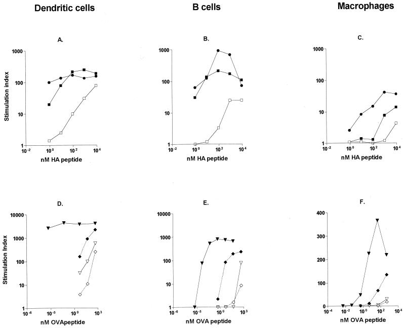
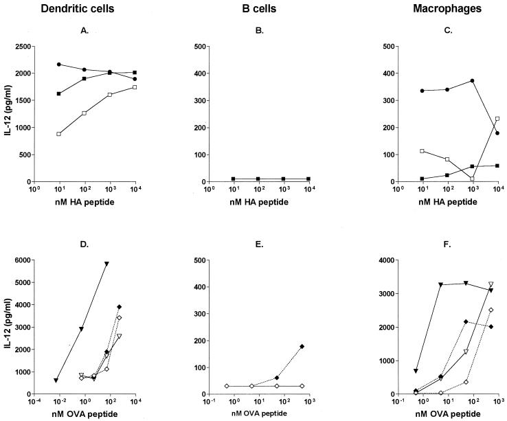
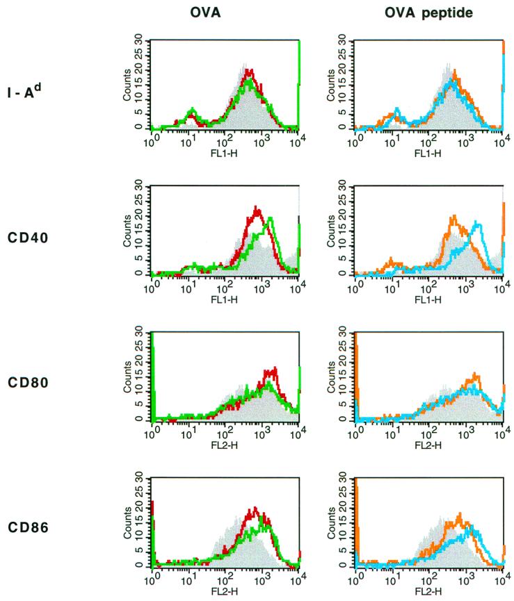
Cholera toxin B subunit (CTB) is an efficient mucosal carrier molecule for the generation of mucosal antibody responses and/or induction of systemic T-cell tolerance to linked antigens. CTB binds with high affinity to GM1 ganglioside cell surface receptors. In this study, we evaluated how conjugation of a peptide or protein antigen to CTB by chemical coupling or genetic fusion influences the T-cell-activating capacity of different antigen-presenting cell (APC) subsets. Using an in vitro system in which antigen-pulsed APCs were incubated with antigen-specific, T-cell receptor-transgenic T cells, we found that the dose of antigen required for T-cell activation could be decreased >10,000-fold using CTB-conjugated compared to free antigen. In contrast, no beneficial effects were observed when CTB was simply admixed with antigen. CTB conjugation enhanced the antigen-presenting capacity not only of dendritic cells and B cells but also of macrophages, which expressed low levels of cell surface major histocompatibility complex (MHC) class II and were normally poor activators of naive T cells. Enhanced antigen-presenting activity by CTB-linked antigen resulted in both increased T-cell proliferation and increased interleukin-12 and gamma interferon secretion and was associated with up-regulation of CD40 and CD86 on the APC surface. These results imply that conjugation to CTB dramatically lowers the threshold concentration of antigen required for immune cell activation and also permits low-MHC II-expressing APCs to prime for a specific immune response.
Figures
Similar articles
-
Cholera toxin and its B subunit promote dendritic cell vaccination with different influences on Th1 and Th2 development.Infect Immun. 2003 Apr;71(4):1740-7. doi: 10.1128/IAI.71.4.1740-1747.2003. Infect Immun. 2003. PMID: 12654787 Free PMC article.
-
B lymphocytes promote expansion of regulatory T cells in oral tolerance: powerful induction by antigen coupled to cholera toxin B subunit.J Immunol. 2008 Dec 15;181(12):8278-87. doi: 10.4049/jimmunol.181.12.8278. J Immunol. 2008. PMID: 19050244
-
Cholera toxin B subunit binding to an antigen-presenting cell directly co-stimulates cytokine production from a T cell clone.Int Immunol. 1996 Dec;8(12):1849-56. doi: 10.1093/intimm/8.12.1849. Int Immunol. 1996. PMID: 8982769
-
ADP-ribosylating bacterial enzymes for the targeted control of mucosal tolerance and immunity.Ann N Y Acad Sci. 2004 Dec;1029:193-208. doi: 10.1196/annals.1309.036. Ann N Y Acad Sci. 2004. PMID: 15681758 Review.
-
Strategies for the induction of immune responses at mucosal surfaces making use of cholera toxin B subunit as immunogen, carrier, and adjuvant.Am J Trop Med Hyg. 1994;50(5 Suppl):42-54. Am J Trop Med Hyg. 1994. PMID: 8203723 Review.
Cited by
-
Protective immunity against Naegleria fowleri infection on mice immunized with the rNfa1 protein using mucosal adjuvants.Parasitol Res. 2015 Apr;114(4):1377-85. doi: 10.1007/s00436-015-4316-3. Epub 2015 Jan 22. Parasitol Res. 2015. PMID: 25604672
-
Assembly of cholera toxin B subunit full-length rotavirus NSP4 fusion protein oligomers in transgenic potato.Plant Cell Rep. 2003 Jun;21(9):884-90. doi: 10.1007/s00299-003-0599-4. Epub 2003 Mar 22. Plant Cell Rep. 2003. PMID: 12789506
-
Transcytosis-blocking abs elicited by an oligomeric immunogen based on the membrane proximal region of HIV-1 gp41 target non-neutralizing epitopes.Curr HIV Res. 2008 May;6(3):218-29. doi: 10.2174/157016208784324994. Curr HIV Res. 2008. PMID: 18473785 Free PMC article.
-
A Vibrio cholerae ghost-based subunit vaccine induces cross-protective chlamydial immunity that is enhanced by CTA2B, the nontoxic derivative of cholera toxin.FEMS Immunol Med Microbiol. 2009 Mar;55(2):280-91. doi: 10.1111/j.1574-695X.2008.00493.x. Epub 2008 Nov 18. FEMS Immunol Med Microbiol. 2009. PMID: 19040663 Free PMC article.
-
Enhanced antigen uptake by dendritic cells induced by the B pentamer of the type II heat-labile enterotoxin LT-IIa requires engagement of TLR2.Vaccine. 2010 May 7;28(21):3696-705. doi: 10.1016/j.vaccine.2010.03.016. Epub 2010 Mar 21. Vaccine. 2010. PMID: 20332049 Free PMC article.
References
-
- Ågren L C, Ekman L, Löwenadler B, Lycke N Y. A genetically engineered nontoxic vaccine adjuvant that combines B cell targeting with immunomodulation by cholera toxin A1 subunit. J Immunol. 1997;158:3936–3946. - PubMed
-
- Arakawa T, Yu J, Chong D K, Hough J, Engen P C, Langridge W H. A plant-based cholera toxin B subunit-insulin fusion protein protects against the development of autoimmune diabetes. Nat Biotechnol. 1998;16:934–938. - PubMed
-
- Bäckström M, Holmgren J, Schödel F, Lebens M. Characterization of an internal permissive site in the cholera toxin B-subunit and insertion of epitopes from human immunodeficiency virus-1, hepatitis B virus and enterotoxigenic Escherichia coli. Gene. 1995;165:163–171. - PubMed
-
- Bäckström M, Lebens M, Schödel F, Holmgren J. Insertion of a HIV-1-neutralizing epitope in surface-exposed internal region of the cholera toxin B-subunit. Gene. 1994;149:211–217. - PubMed
-
- Banchereau J, Steinman R M. Dendritic cells and the control of immunity. Nature. 1998;392:245–252. - PubMed
Publication types
MeSH terms
Substances
LinkOut - more resources
Full Text Sources
Other Literature Sources
Research Materials
Miscellaneous
