. 2001 Sep;69(9):5726-35.
doi: 10.1128/IAI.69.9.5726-5735.2001.

In vivo activation of dendritic cells and T cells during Salmonella enterica serovar Typhimurium infection

Affiliations

In vivo activation of dendritic cells and T cells during Salmonella enterica serovar Typhimurium infection

U Yrlid et al. Infect Immun. 2001 Sep.

Abstract

The present study was initiated to gain insight into the interaction between splenic dendritic cells (DC) and Salmonella enterica serovar Typhimurium in vivo. Splenic phagocytic cell populations associated with green fluorescent protein (GFP)-expressing bacteria and the bacterium-specific T-cell response were evaluated in mice given S. enterica serovar Typhimurium expressing GFP and ovalbumin. Flow cytometry analysis revealed that GFP-positive splenic DC (CD11c+ major histocompatibility complex class II-positive [MHC-II+] cells) were present following bacterial administration, and confocal microscopy showed that GFP-expressing bacteria were contained within CD11c+ MHC-II+ splenocytes. Furthermore, splenic DC and T cells were activated following Salmonella infection. This was shown by increased surface expression of CD86 and CD40 on CD11c+ MHC-II+ cells and increased CD44 and CD69 expression on CD4+ and CD8+ T cells. Salmonella-specific gamma interferon (IFN-gamma)-producing cells in both of these T-cell subsets, as well as cytolytic effector cells, were also generated in mice given live bacteria. The frequency of Salmonella-specific CD4+ T cells producing IFN-gamma was greater than that of specific CD8+ T cells producing IFN-gamma in the same infected animal. This supports the argument that the predominant source of IFN-gamma production by cells of the specific immune response is CD4+ T cells. Finally, DC that phagocytosed live or heat-killed Salmonella in vitro primed bacterium-specific IFN-gamma-producing CD4+ and CD8+ T cells as well as cytolytic effector cells following administration into naïve mice. Together these data suggest that DC are involved in priming naïve T cells to Salmonella in vivo.


Figures
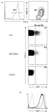
FIG. 1
FIG. 1
Splenic DC associate with GFP-expressing Salmonella. (a) Mice were given either 6 × 10⁸ live or 3 × 10⁹ heat-killed χ4550 OVA-GFP as indicated. Four hours later, splenocytes from two mice were pooled, splenic DC were purified on a MACS column and stained with 7AAD, PE-conjugated anti-CD11c, and biotinylated anti-MHC-II followed by streptavidin-allophycocyanin. The R1 region marked in the histogram indicates the CD11c gate set to analyze MHC-II expression shown in the adjacent contour plot. The contour plot shows the MHC-II expression on the gated CD11c+ splenocytes (R1). The region R2 indicates the CD11c+ MHC-II+ cell population that was analyzed for GFP fluorescence and is shown in the three dot plots. The number in each dot plot represents the percentage of CD11c+ MHC-II+ cells (defined by gates R1 plus R2) that are also GFP+. A total of 200,000 CD11c+ MHC-II+ cells were analyzed for GFP fluorescence. The panel marked control is DC purified from untreated mice. Mice immunized with either live or heat-killed χ4550 OVA resulted in detection of 0.01% GFP+ cells within R2, i.e., the same level as that in control mice. The results are representative of four independent experiments. (b) The histogram shows the GFP fluorescence of χ4550 OVA (dotted line), live χ4550 OVA-GFP (thin line), and heat-killed χ4550 OVA-GFP (thick line). Flow cytometry analysis on live or heat-killed χ4550 OVA-GFP was performed after 4 h in IMDM containing 10% mouse serum at 37°C.
FIG. 2
FIG. 2
GFP+ Salmonella is contained within CD11c+ MHC-II+ splenocytes. Four hours following i.p. administration of 5 × 10⁸ χ4550 OVA-GFP cells, splenocytes from two mice were pooled, and splenic CD11c+ DC were purified on a MACS column and stained with Cy5-conjugated anti-CD11c (blue) and biotinylated anti MHC-II followed by streptavidin-Alexa Flour 594 (red). Cells were subsequently fixed with paraformaldehyde, placed on a polylysine-coated microscope slide, and inspected by epifluorescence and laser confocal microscopy. The left panel shows a section through a CD11c+ MHC-II+ cell containing two GFP+ bacteria. The right panels show the images taken when the cell was cut along the indicated a, b, or c axis and confirms that the GFP+ bacteria remain confined within the CD11c+ MHC-II+ cell surface.
FIG. 3
FIG. 3
Splenic DC are activated in mice given a single dose of S. enterica serovar Typhimurium. Mice were given 2.5 × 10⁶ live or heat-killed χ4550 OVA i.p. Two weeks later, splenocytes from individual mice were stained with 7AAD, FITC-conjugated anti-MHC-II, Cy5-conjugated anti-CD11c, and either PE-conjugated anti-CD86 or anti-CD40 and were analyzed by four-color flow cytometry. The histograms show the CD86 and CD40 expression on gated CD11c+ MHC-II+ splenocytes (R2 as in Fig. 1a) from mice given either live (thick line) or heat-killed (dotted line) χ4550 OVA. The thin line represents the fluorescence intensity of the gated CD11c+ MHC-II+ splenocytes from untreated mice. The staining of cells from an infected mouse using an isotype-matched control antibody is indicated. A similar level of staining using an isotype control antibody was also observed on splenocytes from mice immunized with heat-killed bacteria or naïve mice (data not shown). A total of 10,000 CD11c+ MHC-II+ cells were analyzed for CD86 or CD40 expression. The results shown are from individual mice and are representative of four independent experiments with a total of six mice per group.
FIG. 4
FIG. 4
CD44 and CD69 surface expression is enhanced on CD4+ and CD8+ splenic T cells from mice given a single dose of χ4550 OVA. Mice were given 2.5 × 10⁶ live or heat-killed χ4550 OVA i.p. Two weeks later, splenocytes from individual mice were stained with 7AAD, Allophycocyanin-conjugated TCRαβ, and either FITC-conjugated anti-CD4 or anti-CD8. Cells were subsequently stained with either PE-conjugated anti-CD44 or anti-CD69. The top two histograms show CD44 and CD69 expression on gated TCRαβ+ CD4+ cells, while the bottom histograms show CD44 and CD69 expression on gated TCRαβ+ CD8+ cells from the spleens of mice given either live (thick line) or heat-killed (dotted line) χ4550 OVA. The x axes represent log fluorescence intensity, while the y axes represent the number of gated events as indicated. CD44 and CD69 expression on gated CD4+ and CD8+ T cells from untreated mice (thin line) is shown for comparison. A total of 10,000 gated T cells were analyzed for CD44 or CD69 expression. The results shown are from individual mice and are representative of seven independent experiments with a total of nine mice per group. Appropriate isotype subclass control antibodies showed no significant staining on gated CD4+ and CD8+ splenocytes.
FIG. 5
FIG. 5
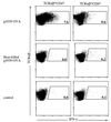
Both CD4+ and CD8+ Salmonella-specific IFN-γ-secreting T cells are elicited in mice given χ4550 OVA. Mice were given either 106 live or heat-killed χ4550 OVA cells or were left untreated (control) as indicated. Two weeks later, splenocytes from individual mice were restimulated in vitro with DC loaded with heat-killed χ4550 OVA for 24 h and were subsequently analyzed for intracellular IFN-γ. Restimulated splenocytes were stained with 7AAD, allophycocyanin-conjugated anti-TCRαβ, and either PE-conjugated anti-CD4 and anti-IFN-γ–FITC or PE-conjugated anti-CD8 and anti-IFN-γ–FITC. Dot plots show IFN-γ-producing CD4+ cells (left) and CD8+ T cells (right). The number in each dot plot represents the percentage of CD4+ or CD8+ T cells staining positive for intracellular IFN-γ in the indicated gates. Splenocytes from untreated (control) mice restimulated in vitro with DC loaded with heat-killed χ4550 OVA had ≤0.1% of gated cells that stained positive for IFN-γ. Likewise, splenocytes from χ4550 OVA-infected mice restimulated in vitro with DC in medium alone stained positive for IFN-γ at a low level (≤ 0.8% of CD4+ or 0.2% CD8+ gated T cells). FITC-labeled isotype-matched control antibodies stained less than 0.1% of the CD4+ or CD8+ T cells (not shown). A total of 30,000 gated T cells were analyzed. The results shown are from individual mice and are representative of four independent experiments with a total of seven mice per group.
FIG. 6
FIG. 6
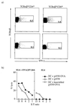
DC loaded with live Salmonella induce bacterium-specific IFN-γ-producing CD4+ and CD8+ T cells and cytotoxic effector cells. (a) Mice were given DC loaded with χ4550 OVA or control DC as indicated on two occasions 1 week apart. Two weeks after the last administration, splenocytes from individual mice were restimulated in vitro with DC loaded with heat-killed χ4550 OVA for 24 h and were subsequently analyzed for intracellular IFN-γ. Restimulated splenocytes were stained with 7AAD, biotinylated anti-CD4 followed by streptavidin-allophycocyanin, PE-conjugated anti-CD8, and anti-IFN-γ–FITC. Dot plots of flow cytometry analysis of CD4+ (left) and CD8+ (right) are shown. The number in each dot plot is the percentage of CD4+ or CD8+ cells staining positive for intracellular IFN-γ in the indicated gates. Splenocytes from mice given control DC restimulated in vitro with DC only stained positive for IFN-γ at a low level (0.1% of CD4+ or CD8+ gated cells). Likewise, splenocytes from mice given DC loaded with χ4550 OVA restimulated in vitro with DC only stained positive for IFN-γ at a low level (≤0.9% of CD4+ or 0.1% CD8+ gated cells). FITC-labeled isotype-matched control antibodies stained less than 0.1% of the cells in the indicated gates. A total of 30,000 gated CD4+ or CD8+ cells were analyzed. (b) Mice were given either DC loaded with live χ4550 OVA (DC + χ4550 OVA), DC loaded with live χ4550 not expressing OVA (DC + χ4550), control DC (DC), or free χ4550 OVA as indicated. Two weeks after the second injection, splenocytes from individual mice were restimulated in vitro with OVA(257-264) peptide and were analyzed for cytotoxic activity in 51Cr release assays. The percent lysis of either OVA(257-264)-loaded (left panels) or unloaded (right panels) EL4 target cells is shown. The results shown are from individual mice and are representative of four independent experiments with a total of four mice per group.
FIG. 7
FIG. 7
DC loaded with heat-killed Salmonella induce bacterium-specific IFN-γ-producing and cytotoxic T cells. (a) Mice were immunized with DC loaded with heat-killed χ4550 OVA on two occasions 1 week apart. Splenocytes from individual mice were restimulated with DC loaded with heat-killed χ4550 OVA (top two dot plots) or with DC only (bottom two dot plots). The cells were then stained with 7AAD and allophycocyanin-conjugated anti-TCRαβ and either PE-conjugated anti-CD4 or anti-CD8 followed by anti-IFN-γ–FITC. Dot plots of flow cytometry analysis of TCRαβ+ CD4+ (left) and TCRαβ+ CD8+ (right) are shown. The number in each dot plot shows the percentage of cells staining positive for intracellular IFN-γ. FITC-labeled isotype-matched control antibodies stained less than 0.1% of the T cells. A total of 80,000 T cells were analyzed. (b) Mice were given DC loaded with either live (DC + χ4550 OVA) or heat-killed (DC + heat-killed χ4550 OVA) χ4550 OVA or with live χ4550 not expressing OVA (DC + χ4550) as indicated. Cytotoxic activity was measured as in Fig. 6b. The results shown are from individual mice and are representative of four independent experiments with a total of four mice per group.

Similar articles

Cited by

References

    1. Austyn J M, Gordon S. F4/80, a monoclonal antibody directed specificially against the mouse macrophage. Eur J Immunol. 1981;11:805–815. - PubMed
    1. Banchereau J, Briere F, Caux C, Davoust J, Lebecque S, Liu Y J, Pulendran B, Palucka K. Immunobiology of dendritic cells. Annu Rev Immunol. 2000;18:767–811. - PubMed
    1. Banchereau J, Steinman R M. Dendritic cells and the control of immunity. Nature. 1998;392:245–252. - PubMed
    1. Buchmeier N A, Heffron F. Intracellular survival of wild-type Salmonella typhimurium and macrophage-sensitive mutants in diverse populations of macrophages. Infect Immun. 1989;57:1–7. - PMC - PubMed
    1. Cobbold S P, Jayasuriya A, Nash A, Prospero T D, Waldmann H. Therapy with monoclonal antibodies by elimination of T-cell subsets in vivo. Nature. 1984;312:548–551. - PubMed

Publication types

MeSH terms