

Type 3 fimbrial shaft (MrkA) of Klebsiella pneumoniae, but not the fimbrial adhesin (MrkD), facilitates biofilm formation
- PMID: 11500458
- PMCID: PMC98698
- DOI: 10.1128/IAI.69.9.5805-5812.2001
Type 3 fimbrial shaft (MrkA) of Klebsiella pneumoniae, but not the fimbrial adhesin (MrkD), facilitates biofilm formation
Abstract
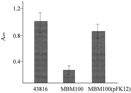
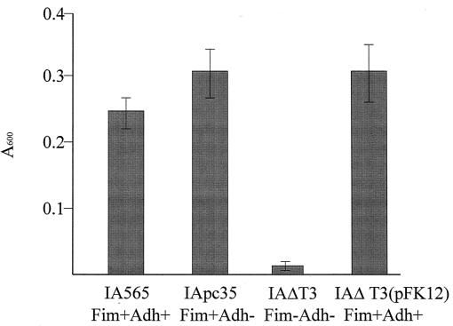
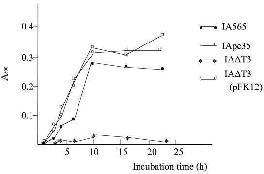
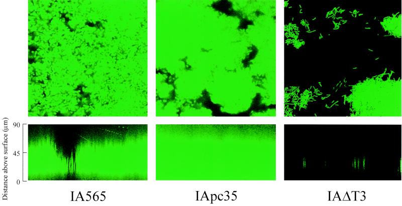
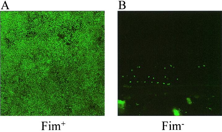
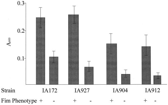
Isolates of Klebsiella pneumoniae are responsible for opportunistic infections, particularly of the urinary tract and respiratory tract, in humans. These bacteria express type 3 fimbriae that have been implicated in binding to eucaryotic cells and matrix proteins. The type 3 fimbriae mediate binding to target tissue using the MrkD adhesin that is associated with the fimbrial shaft comprised of the MrkA protein. The formation of biofilms in vitro by strains of K. pneumoniae was shown to be affected by the production of fimbriae on the bacterial surface. However, a functional MrkD adhesin was not necessary for efficient biofilm formation. Nonfimbriate strains were impaired in their ability to form biofilms. Using isogenic fimbriate and nonfimbriate strains of K. pneumoniae expressing green fluorescent protein it was possible to demonstrate that the presence of type 3 fimbriae facilitated the formation of dense biofilms in a continuous-flowthrough chamber. Transformation of nonfimbriate mutants with a plasmid possessing an intact mrk gene cluster restored the fimbrial phenotype and the rapid ability to form biofilms.
Figures
Similar articles
-
Klebsiella pneumoniae MrkD-mediated biofilm formation on extracellular matrix- and collagen-coated surfaces.Microbiology (Reading). 2003 Sep;149(Pt 9):2397-2405. doi: 10.1099/mic.0.26434-0. Microbiology (Reading). 2003. PMID: 12949165
-
Role of type 1 and type 3 fimbriae in Klebsiella pneumoniae biofilm formation.BMC Microbiol. 2010 Jun 23;10:179. doi: 10.1186/1471-2180-10-179. BMC Microbiol. 2010. PMID: 20573190 Free PMC article.
-
Adherence properties of an mrkD-negative mutant of Klebsiella pneumoniae.Infect Immun. 1995 May;63(5):2026-32. doi: 10.1128/iai.63.5.2026-2032.1995. Infect Immun. 1995. PMID: 7729917 Free PMC article.
-
Klebsiella pneumoniae and type 3 fimbriae: nosocomial infection, regulation and biofilm formation.Future Microbiol. 2012 Aug;7(8):991-1002. doi: 10.2217/fmb.12.74. Future Microbiol. 2012. PMID: 22913357 Review.
-
Combining sites of bacterial fimbriae.Curr Opin Struct Biol. 2007 Oct;17(5):506-12. doi: 10.1016/j.sbi.2007.06.011. Epub 2007 Aug 23. Curr Opin Struct Biol. 2007. PMID: 17719218 Review.
Cited by
-
Preventing dysbiosis of the neonatal mouse intestinal microbiome protects against late-onset sepsis.Nat Med. 2019 Nov;25(11):1772-1782. doi: 10.1038/s41591-019-0640-y. Epub 2019 Nov 7. Nat Med. 2019. PMID: 31700190 Free PMC article.
-
The KP1_4563 gene is regulated by the cAMP receptor protein and controls type 3 fimbrial function in Klebsiella pneumoniae NTUH-K2044.PLoS One. 2017 Jul 21;12(7):e0180666. doi: 10.1371/journal.pone.0180666. eCollection 2017. PLoS One. 2017. PMID: 28732013 Free PMC article.
-
1H, 13C and 15N assignment of self-complemented MrkA protein antigen from Klebsiella pneumoniae.Biomol NMR Assign. 2024 Dec;18(2):171-179. doi: 10.1007/s12104-024-10185-3. Epub 2024 Jul 17. Biomol NMR Assign. 2024. PMID: 39018011 Free PMC article.
-
Biofilm formed by a hypervirulent (hypermucoviscous) variant of Klebsiella pneumoniae does not enhance serum resistance or survival in an in vivo abscess model.Virulence. 2012 May 1;3(3):309-18. doi: 10.4161/viru.20383. Epub 2012 May 1. Virulence. 2012. PMID: 22546898 Free PMC article.
-
Genome-scale metabolic modeling reveals increased reliance on valine catabolism in clinical isolates of Klebsiella pneumoniae.NPJ Syst Biol Appl. 2022 Oct 28;8(1):41. doi: 10.1038/s41540-022-00252-7. NPJ Syst Biol Appl. 2022. PMID: 36307414 Free PMC article.
References
-
- Adegbola R A, Old D C. Fimbrial haemagglutinins in Enterobacter species. J Gen Microbiol. 1983;129:2175–2180. - PubMed
-
- Clegg S, Korhonen K T, Hornick B D, Tarkkanen A-M. Type 3 fimbriae of the Enterobacteriaceae. In: Klemm P, editor. Fimbriae: adhesion, genetics, biogenesis, and vaccines. Boca Raton, Fla: CRC Press; 1994. pp. 97–104.
-
- Costerton J W, Stewart P S, Greenberg E P. Bacterial biofilms: a common cause of persistent infections. Science. 1999;284:1318. - PubMed
-
- Costerton J W, Cheng K-J, Geesey G G, Ladd T I, Nickel J C, Dasgupta M, Marrie T J. Bacterial biofilms in nature and disease. Annu Rev Microbiol. 1987;41:435–464. - PubMed
Publication types
MeSH terms
Substances
Grants and funding
LinkOut - more resources
Full Text Sources
Other Literature Sources
Molecular Biology Databases
