. 2001 Sep;69(9):5899-904.
doi: 10.1128/IAI.69.9.5899-5904.2001.

Isolation and characterization of a mutant Chinese hamster ovary cell line that is resistant to Chlamydia trachomatis infection at a novel step in the attachment process

Affiliations

Isolation and characterization of a mutant Chinese hamster ovary cell line that is resistant to Chlamydia trachomatis infection at a novel step in the attachment process

R A Carabeo et al. Infect Immun. 2001 Sep.

Abstract

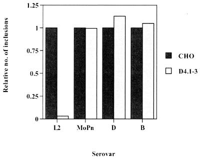
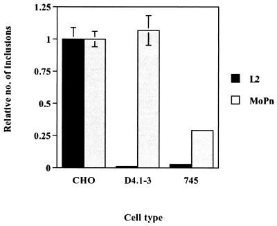
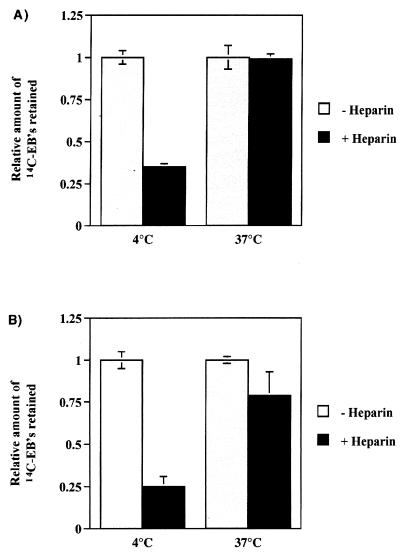
Host factors involved in Chlamydia trachomatis pathogenesis were investigated by random chemical mutagenesis of Chinese hamster ovary (CHO-K1) cells followed by selection for clones resistant to chlamydial infection. A clonal mutant cell line, D4.1-3, refractory to infection by the C. trachomatis L2 serovar was isolated. The D4.1-3 cell line appears to be lacking in a previously undescribed temperature-dependent and heparin-resistant binding step that occurs subsequent to engagement of cell surface heparan sulfate by L2 elementary bodies. This novel binding step differentiates the lymphogranuloma venereum (LGV) serovar from other serovars and may contribute the different pathologies associated with LGV and non-LGV strains.


Figures

FIG. 1
FIG. 1
Susceptibilities of the D4.1–3 mutant cell line to infection by serovars L2, MoPn, B, and D. Cells were preincubated in 42 μg of DEAE-dextran/ml in minimal essential medium for 20 min at 37°C, inoculated with Renografin density gradient-purified (5) L2, MoPn, B, and D EBs (MOI = 0.5) at 37°C for 1 h, washed three times with heparin to remove cell surface-associated EBs, and incubated at 37°C for 24 h. The inclusion-forming assay was performed essentially as described by Furness et al. (10). At 24 h postinfection, infected cells were methanol fixed and processed for immunofluorescence microscopy, using a rabbit polyclonal anti-C. trachomatis L2 EB antibody and a fluorescein isothiocyanate-conjugated anti-rabbit immunoglobulin (Zymed, South San Francisco, Calif.). Data are from triplicates and represented as mean ± standard deviation.
FIG. 2
FIG. 2
Immunofluorescence images of parental CHO-K1 and D4.1–3 cell lines infected with serovar L2 or serovar D EBs. Cells were inoculated with serovar L2 or D EBs (MOI = 500) at 4°C for 1 h. The cells were washed with cold 1× HBSS three times and were immediately incubated at 37°C for 10 min. The cells were rinsed three times with heparin and fixed with methanol for immunofluorescence microscopy. (A) CHO infected with L2; (B) D4.1–3 infected with L2; (C) CHO infected with D; and (D) D4.1–3 infected with D.
FIG. 3
FIG. 3
Differential susceptibility of D4.1–3 mutant cells and GAG-deficient pgsA-745 cells to infection by C. trachomatis serovar MoPn EBs. Cells were inoculated with L2 or MoPn EBs (MOI = 0.5) for 1 h at 37°C. The cells were rinsed three times with HBSS and were incubated at 37°C for 24 h. Inclusions were visualized by immunofluorescence microscopy and counted. Data are from triplicates and are represented as mean ± standard deviation.
FIG. 4
FIG. 4
Effects of incubation temperature and heparin treatment on the attachment of 14C-labeled EBs to live (A) or paraformaldehyde-fixed (B) CHO-K1 cells. Cells were infected with 14C-labeled EBs (MOI = 500) for 30 min at either 4 or 37°C. Quality of binding was assessed by release with HBSS or heparin washes. Retained radioactivity was counted by scintillation counting. Data are from triplicates and are represented as mean ± standard deviation.
FIG. 5
FIG. 5
Kinetics of binding of 14C-labeled L2 EBs to paraformaldehyde-fixed CHO, D4.1–3, and pgsA-745 cells. 14C-labeled L2 EBs in 200 μl of HBSS (≈2.4 × 10⁴ cpm) were added to each well. The radioactive inoculum was removed at the indicated times after incubation at 37°C, and the cells were rinsed three times, either with HBSS alone (A) or supplemented with 2 μg of heparin/ml (B) (USB, Cleveland, Ohio). The fixed cells were solubilized with 2% sodium dodecyl sulfate in phosphate-buffered saline, and associated radioactivity was monitored by scintillation counting. The data was plotted using Cricket Graph version 1.5.1 Data are from triplicates and are represented as mean ± standard deviation.

References

    1. Bair C-H, Chung C-S, Vasilevskaya I A, Chang W. Isolation and characterization of a Chinese hamster ovary mutant cell line with altered sensitivity to vaccinia virus killing. J Virol. 1996;70:4655–4666. - PMC - PubMed
    1. Becker Y, Hochberg E, Zakay-Rones Z. Interaction of trachoma elementary bodies with host cells. Isr J Med Sci. 1969;5:121–124. - PubMed
    1. Bose S K, Paul R G. Purification of Chlamydia trachomatis lymphogranuloma venereum elementary bodies and their interaction with HeLa cells. J Gen Microbiol. 1982;128:1371–1379. - PubMed
    1. Boutin E L, Sanderson R D, Bernfield M, Cunha G R. Epithelial-mesenchymal interactions in uterus and vagina alter the expression of the cell surface proteoglycan, syndecan. Dev Biol. 1991;143:63–74. - PubMed
    1. Caldwell H D, Kromhout J, Schachter J. Purification and partial characterization of the major outer membrane protein of Chlamydia trachomatis. Infect Immun. 1981;31:1161–1176. - PMC - PubMed

LinkOut - more resources