Comparative Study
. 2001 Aug;126(4):1370-80.
doi: 10.1104/pp.126.4.1370.

Overexpression of KNAT1 in lettuce shifts leaf determinate growth to a shoot-like indeterminate growth associated with an accumulation of isopentenyl-type cytokinins

Affiliations
Comparative Study

Overexpression of KNAT1 in lettuce shifts leaf determinate growth to a shoot-like indeterminate growth associated with an accumulation of isopentenyl-type cytokinins

G Frugis et al. Plant Physiol. 2001 Aug.

Abstract

Leaves are specialized organs characterized by defined developmental destiny and determinate growth. The overexpression of Knotted1-like homeobox genes in different species has been shown to alter leaf shape and development, but a definite role for this class of genes remains to be established. Transgenics that overexpress Knotted1-like genes present some traits that are characteristic of altered cytokinin physiology. Here we show that lettuce (Lactuca sativa) leaves that overexpress KNAT1, an Arabidopsis kn1-like gene, acquire characteristics of indeterminate growth typical of the shoot and that this cell fate change is associated with the accumulation of specific types of cytokinins. The possibility that the phenotypic effects of KNAT1 overexpression may arise primarily from the modulation of local ratios of different cytokinins is discussed.


Figures
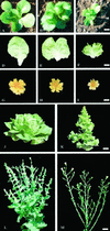
Figure 1
Figure 1
Phenotypical alterations of PetE::KNAT1 lettuce T1 plants. PetE::KNAT1 T1 plants were placed in three phenotypic categories based on leaf morphology. A, D, and G, Normal, plants that fully resembled wild-type phenotype. B, E, and H, Mild, plants that resemble the parental phenotype. C, F, and I, Severe, plants that show severe alterations of leaf shape and plant architecture. A through C, Six-leaves-stage plants grown in greenhouse; D through F, 10th leaf of same age from wild-type, mild, and severe PetE::KNAT1 T1 plants, respectively; G through I, inflorescence from wild-type, mild, and severe PetE::KNAT1 T1 plants, respectively; wild-type (J) and severe transgenic (K) adult plants of same age. Transgenic lettuce always flowers several days earlier than wild-type plants. Wild-type (L) and severe transgenic (M) plants at flowering time. PetE::KNAT1 inflorescence stem displays loss of apical dominance with formation of several floral branches of different length with respect to the more regular architecture of the wild-type. Size bar in C for A through C = 1 cm. Size bar in F for D through F = 1.2 cm. Size bar in I for G through I = 0.7 cm. Size bar in K for J and K = 2.5 cm. Size bar in M for L and M = 1.5 cm.
Figure 2
Figure 2
Alteration of PetE::KNAT1 leaf shape and venation pattern at different developmental stages. A, One-leaf stage plants from lettuce wild-type and T2 PetE::KNAT1. Size bar in M = 0.3 cm. B through L, Embossed modified images of cleared leaves: first leaf from wild-type and T2 PetE::KNAT1 with the characteristic heart-shape due to a reduced elongation of the midvein; size bar in M = 1 mm (B); eighth leaf in expansion phase from wild-type and T2 PetE::KNAT1; size bar in M = 0.5 cm (C); D through I, detail of F and G leaf tip, H and I leaf base, and H and I midvein from which secondary veins depart from wild type (D, F, and H) and PetE::KNAT1 (E, G, and I). Size bar in M: for D and E = 90 μm, for F and G = 100 μm, and for H and I = 50 mm. J through M, Pattern of leaf-like structures formation from the PetE:KNAT1 leaf margins: sequential images (embossed modified images from cleared leaves) of outcoming structures that arise from PetE:KNAT1 leaf margin tissues. The first step involves the elongation of a single vein (J) that protrudes from the leaf edge in correspondence of groups of cells with meristematic features. The main vein subsequently branches (K) to form secondary strands that eventually protrude out from the main structure and give rise to leaf-like tissues in a reiterative process of morphogenesis (L). Size bar in M = 0.12 cm. M, Advanced leaf-like structures at the PetE::KNAT1 leaf margin; size bar = 0.2 cm.
Figure 3
Figure 3
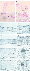
Histological characterization of PetE::KNAT1 leaves. A, Transverse section (treated by the Feulgen reaction that stains only DNA) of PetE::KNAT1 leaf margin in correspondence to neoforming leaf-like structures. Size bar = 50 μm. B, Magnification of section in A. Cells present high nucleus/cytoplasm ratio and mitotic figures. Arrow indicates an example of cells in mitotic phase. Size bar = 40 μm. C, Transverse section of vascular bundles in inner sectors of a PetE::KNAT1 leaf. Arrow indicates interfascicular region between the two bundles. Size bar = 100 μm. D, Magnification of section in C. Arrow indicates elongated meristematic cells typical of cambium initiation tissues in the bundle between differentiated xylem and phloem elements. Size bar = 50 μm. E through L, Comparison of same leaf sectors from wild-type and PetE::KNAT1 plants of same age and same developmental stage. Light microscopy. E, Transverse section of PetE::KNAT1 leaf in close proximity to the margin. The mesophyll thickness is irregular, palisade cells are absent, and intercellular space is rare. F, Detail of a vascular bundle in transverse section of PetE::KNAT1 leaf in close proximity to the margin: irregular shape due to an anomalous and disorganized association of xylem and phloem elements. G, Transverse section of wild-type leaf in close proximity to the margin. Palisade cells are detectable and intercellular space is present. H, Detail of a vascular bundle in transverse section of wild-type leaf in close proximity to the margin. Regular association of xylem and phloem elements. I, Inner sector of PetE::KNAT1 leaf in close proximity to the midvein. The mesophyll is compact, palisade cells are absent, and intercellular space is rare. J, Detail of a vascular bundle in inner sectors of PetE::KNAT1 leaf in close proximity to the midvein: elongated cells typical of cambium initiation tissues are present inside the bundles between differentiated phloem and xylem elements regularly associated. K, Transverse section of inner sectors of wild-type leaf in close proximity to the midvein. Palisade tissue is fully developed and intercellular spaces are present. L, Detail of a vascular bundle in inner sectors of wild-type leaf in close proximity to the midvein. Phloem and xylem are regularly associated and cambial initial cells are never observed in wild-type leaves. Size bar in K for E through K = 60 μm. Size bar in L for F through L = 30 μm.
Figure 4
Figure 4
RNA gel-blot analysis of PetE::KNAT1 transgenic lines. A, Total RNA was isolated from rosette leaves of wild-type cv Luxor lettuce or T1 PetE::KNAT1 transformants with mild or severe leaf phenotype from two independent lines (p173 and p177). Ten micrograms of RNA was loaded per lane and probed with KNAT1 cDNA. Ethidium bromide staining is shown as control for loading. B, Total RNA from different tissues of PetE::KNAT1 homozygous 12-leaves-stage plants (first six lanes) or 4-leaves-stage plants (last three lanes). Ten micrograms of RNA was loaded per lane and probed with KNAT1 cDNA. 5S RNA was used as control for loading.
Figure 5
Figure 5
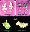
Altered response to hormones of PetE::KNAT1 leaf explants. Leaf explants from wild-type lettuce and PetE::KNAT1 T2 homozygous plants were cultivated in vitro for 30 d in the absence of hormones (top) or in the presence of 1 mg L−1 NAA for 10 d. After 30 d without hormones, PetE::KNAT1 explants are still bright green, whereas wild-type leaves underwent to complete necrosis. In the presence of auxin, PetE::KNAT1 explants completely transform into callus, whereas wild-type leaves give rise exclusively to roots after 10 d.
Figure 6
Figure 6
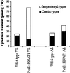
Analysis of cytokinin content in PetE::KNAT1young and adult leaves. Z-type (Z, ZR, DHZ, and DHZR) and isopentenyl-type (IP and IPA) cytokinin content was measured in leaves from adult and young leaves of wild-type and PetE::KNAT1 plants (graphic version of the data presented in Table II).
Figure 7
Figure 7
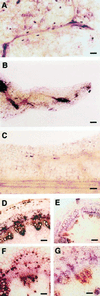
Immunocytolocalization of the cytokinins IP in PetE::KNAT1 leaves and stem. Affinity-purified rabbit antisera were used for immunolocalization of IP in aldehyde-fixed tissues of expanding leaves from PetE:KNAT1 and wild-type lettuce plants. Samples were mounted in a phosphate-buffered saline (PBS)/glycerine mixture (1:1, v/v) and were immediately observed under a photomicroscope (Leica). A, PetE:KNAT1 leaf vascular strands close to the leaf margin. IP strongly accumulate at the vein branching point, in parenchymal cells associated with vascular strands, and in the numerous secretory cells that are present in transgenic leaves (dark elliptical dots in the figure). Size bar = 100 μm. B, PetE:KNAT1 outgrowing structure that forms from the leaf margin. IP accumulates in the vascular bundles at the base of the neoforming structures. Size bar = 100 μm. C, Longitudinal section of a wild-type leaf margin. IP is barely detectable in every tissue. Size bar = 60 μm. D, Transverse section of PetE:KNAT1 stem proximal to the shoot apex. IP is strongly accumulated in vascular bundles. Size bar = 50 μm. E, Transverse section of wild-type stem proximal to the shoot apex. IP is barely detectable in vascular tissues. Size bar = 200 μm. F, Transverse section of PetE:KNAT1 stem proximal to the transition zone between shoot and root. IP is still high in vascular bundles elements. Size bar = 25 μm. G, Transverse section of wild-type stem proximal to the transition zone between shoot and root. IP is still barely detectable in vascular tissues. Size bar = 25 μm.

Similar articles

Cited by

References

    1. Chan RL, Gago GM, Palena CM, Gonzalez DH. Homeoboxes in plant development. Biochim Biophys Acta. 1998;1442:1–19. - PubMed
    1. Chuck G, Lincoln C, Hake S. KNAT1 induces lobed leaves with ectopic meristems when overexpressed in Arabidopsis. Plant Cell. 1996;8:1277–1289. - PMC - PubMed
    1. Coenen C, Lomax TL. Auxin-cytokinin interactions in higher plants: old problems and new tools. Trends Plant Sci. 1997;9:351–356. - PubMed
    1. Curtis IS, Power JB, Blackhall NW, de Laat AMM, Davey MR. Genotype-independent transformation of lettuce using Agrobacterium tumefaciens. J Exp Bot. 1994;279:1441–1449.
    1. Dewitte W, Chiappetta A, Azmi A, Witters E, Strnad M, Rembur J, Noin M, Chriqui D, Van Onckelen H. Dynamics of cytokinins in apical shoot meristems of a day-neutral tobacco during floral transition and flower formation. Plant Physiol. 1999;119:111–122. - PMC - PubMed

Publication types

MeSH terms