

Isolation and characterization of kinase interacting protein 1, a pollen protein that interacts with the kinase domain of PRK1, a receptor-like kinase of petunia
- PMID: 11500547
- PMCID: PMC117148
- DOI: 10.1104/pp.126.4.1480
Isolation and characterization of kinase interacting protein 1, a pollen protein that interacts with the kinase domain of PRK1, a receptor-like kinase of petunia
Abstract
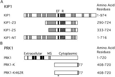
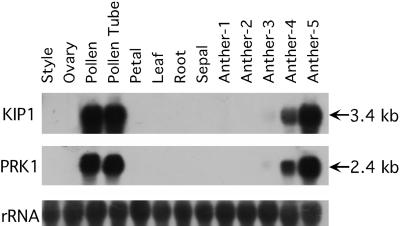
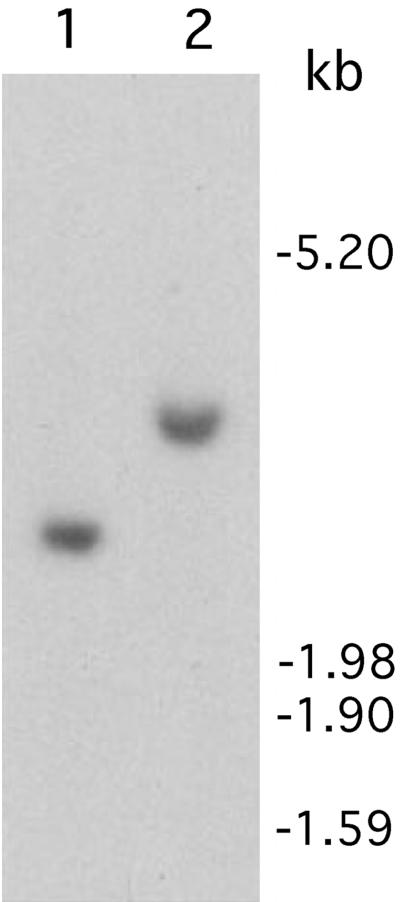
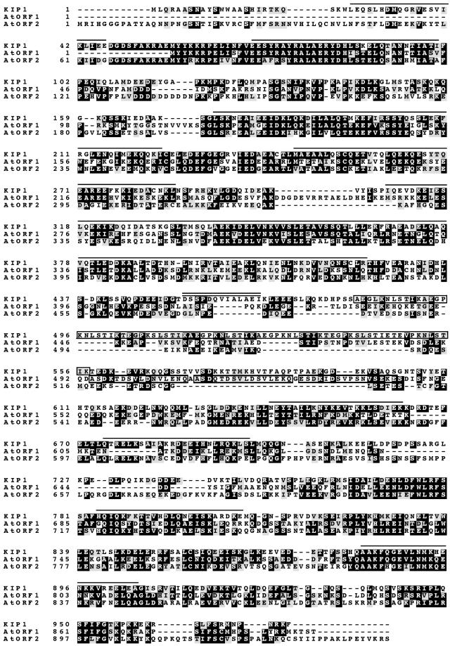
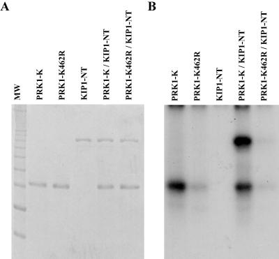
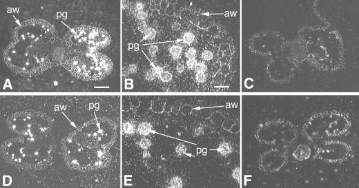
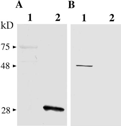
Many receptor-like kinases have been identified in plants and have been shown by genetic or transgenic knockouts to play diverse physiological roles; however, to date, the cytosolic interacting proteins of relatively few of these kinases have been identified. We have previously identified a predominantly pollen-expressed receptor-like kinase of petunia (Petunia inflata), named PRK1, and we have shown by the antisense RNA approach that it is required for microspores to progress from the unicellular to bicellular stage. To investigate the PRK1-mediated signal transduction pathway, PRK1-K cDNA, encoding most of the cytoplasmic domain of PRK1, was used as bait in yeast (Saccharomyces cerevisiae) two-hybrid screens of pollen/pollen tube cDNA libraries of petunia. A protein named kinase interacting protein 1 (KIP1) was found to interact very strongly with PRK1-K. This interaction was greatly reduced when lysine-462 of PRK1-K, believed to be essential for kinase activity, was replaced with arginine (the resulting protein is named PRK1-K462R). The amino acid sequence of KIP1 deduced from full-length cDNA contains an EF-hand Ca(2+)-binding motif and nine predicted coiled-coil regions. The yeast two-hybrid assay and affinity chromatography showed that KIP1 interacts with itself to form a dimer or higher multimer. KIP1 is present in a single copy in the genome, and is expressed predominantly in pollen with a similar temporal pattern to PRK1. In situ hybridization showed that PRK1 and KIP1 transcripts were localized in the cytoplasm of pollen. PRK1-K phosphorylated KIP1-NT (amino acids 1--716), whereas PRK1-K462R only weakly phosphorylated KIP1-NT in vitro.
Figures
Similar articles
-
Identification and characterization of PiORP1, a Petunia oxysterol-binding-protein related protein involved in receptor-kinase mediated signaling in pollen, and analysis of the ORP gene family in Arabidopsis.Plant Mol Biol. 2006 Jul;61(4-5):553-65. doi: 10.1007/s11103-006-0030-y. Plant Mol Biol. 2006. PMID: 16897474
-
Characterization of a pollen-expressed receptor-like kinase gene of Petunia inflata and the activity of its encoded kinase.Plant Cell. 1994 May;6(5):709-21. doi: 10.1105/tpc.6.5.709. Plant Cell. 1994. PMID: 8038606 Free PMC article.
-
Interaction of PRK1 receptor-like kinase with a putative elF2B beta-subunit in tobacco.Mol Cells. 2000 Dec 31;10(6):626-32. doi: 10.1007/s10059-000-0626-z. Mol Cells. 2000. PMID: 11211866
-
Self-incompatibility in Petunia: a self/nonself-recognition mechanism employing S-locus F-box proteins and S-RNase to prevent inbreeding.Wiley Interdiscip Rev Dev Biol. 2012 Mar-Apr;1(2):267-75. doi: 10.1002/wdev.10. Epub 2011 Nov 17. Wiley Interdiscip Rev Dev Biol. 2012. PMID: 23801440 Review.
-
The Ark1/Prk1 family of protein kinases. Regulators of endocytosis and the actin skeleton.EMBO Rep. 2003 Mar;4(3):246-51. doi: 10.1038/sj.embor.embor776. EMBO Rep. 2003. PMID: 12634840 Free PMC article. Review.
Cited by
-
Genetic mapping and molecular characterization of the self-incompatibility (S) locus in Petunia inflata.Plant Mol Biol. 2003 Nov;53(4):565-80. doi: 10.1023/B:PLAN.0000019068.00034.09. Plant Mol Biol. 2003. PMID: 15010619
-
Coiled-coil protein composition of 22 proteomes--differences and common themes in subcellular infrastructure and traffic control.BMC Evol Biol. 2005 Nov 16;5:66. doi: 10.1186/1471-2148-5-66. BMC Evol Biol. 2005. PMID: 16288662 Free PMC article.
-
Identification and characterization of PiORP1, a Petunia oxysterol-binding-protein related protein involved in receptor-kinase mediated signaling in pollen, and analysis of the ORP gene family in Arabidopsis.Plant Mol Biol. 2006 Jul;61(4-5):553-65. doi: 10.1007/s11103-006-0030-y. Plant Mol Biol. 2006. PMID: 16897474
-
Hairpin in a haystack: In silico identification and characterization of plant-conserved microRNA in Rafflesiaceae.Open Life Sci. 2025 Jan 27;20(1):20221033. doi: 10.1515/biol-2022-1033. eCollection 2025. Open Life Sci. 2025. PMID: 39881826 Free PMC article.
-
The Rice DNA-Binding Protein ZBED Controls Stress Regulators and Maintains Disease Resistance After a Mild Drought.Front Plant Sci. 2020 Aug 18;11:1265. doi: 10.3389/fpls.2020.01265. eCollection 2020. Front Plant Sci. 2020. PMID: 33013945 Free PMC article.
References
-
- Ai Y, Singh A, Coleman CE, Ioerger TR, Kheyr-Pour A, Kao T-h. Self-incompatibility in Petunia inflata: isolation and characterization of cDNAs encoding three S allele-associated proteins. Sex Plant Reprod. 1990;3:130–138.
-
- Ausubel FM, Brent R, Kingston RE, Moore DD, Seidman JG, Smith JA, Struhl K. Current Protocols in Molecular Biology. Vol. 2. New York: Wiley Interscience; 1993. pp. 13.13.5–13.13.9.
-
- Becraft PW. Receptor kinases in plant development. Trends Plant Sci. 1998;3:384–388.
-
- Braun DM, Stone JM, Walker JC. Interaction of the maize and Arabidopsis kinase interaction domain with a subset of receptor-like protein kinases: implication for transmembrane signaling in plants. Plant J. 1997;12:83–95. - PubMed
Publication types
MeSH terms
Substances
Associated data
- Actions
LinkOut - more resources
Full Text Sources
Research Materials
Miscellaneous
