Comparative Study
. 2001 Aug 20;154(4):731-40.
doi: 10.1083/jcb.200104045. Epub 2001 Aug 13.

The discrepancy between presenilin subcellular localization and gamma-secretase processing of amyloid precursor protein

Affiliations
Comparative Study

The discrepancy between presenilin subcellular localization and gamma-secretase processing of amyloid precursor protein

P Cupers et al. J Cell Biol. .

Abstract

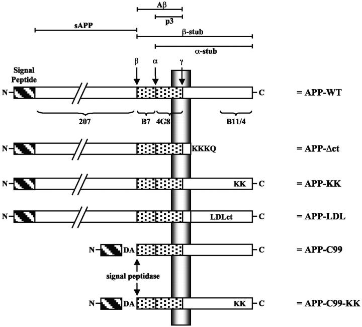
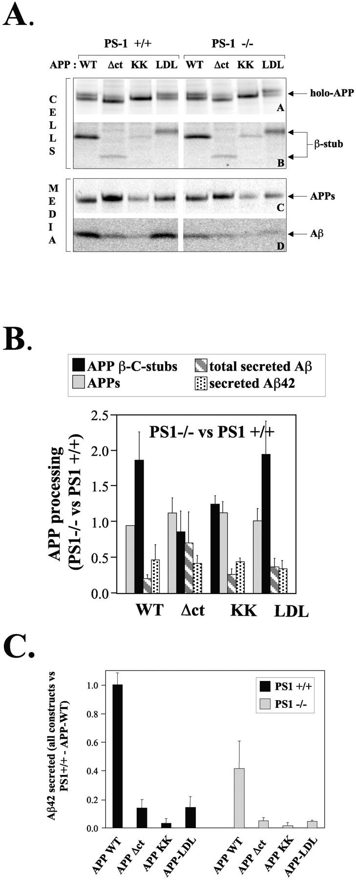
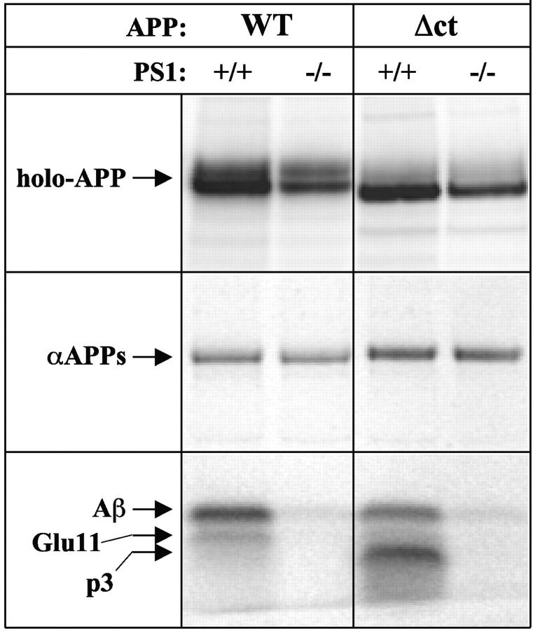
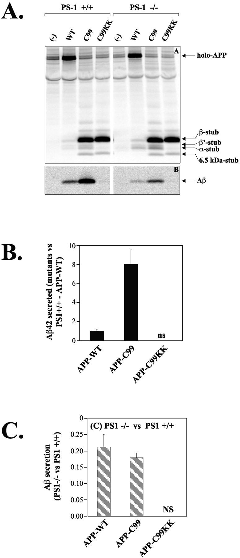
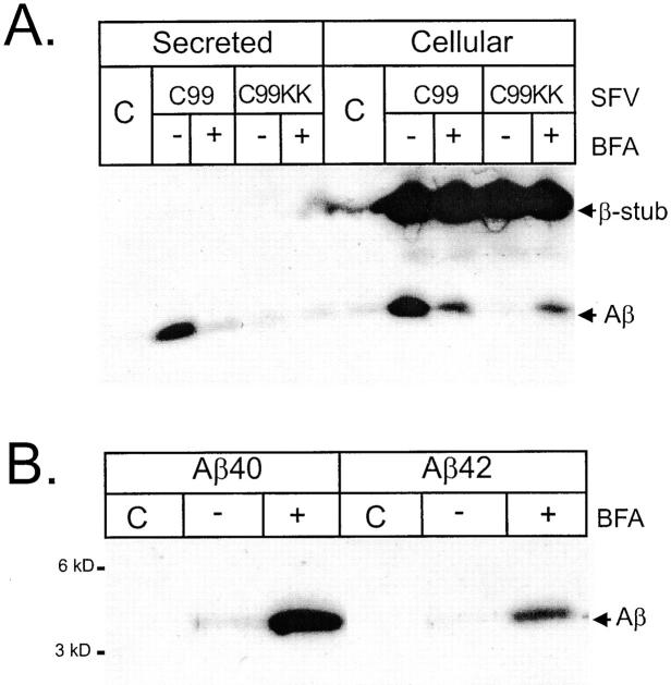
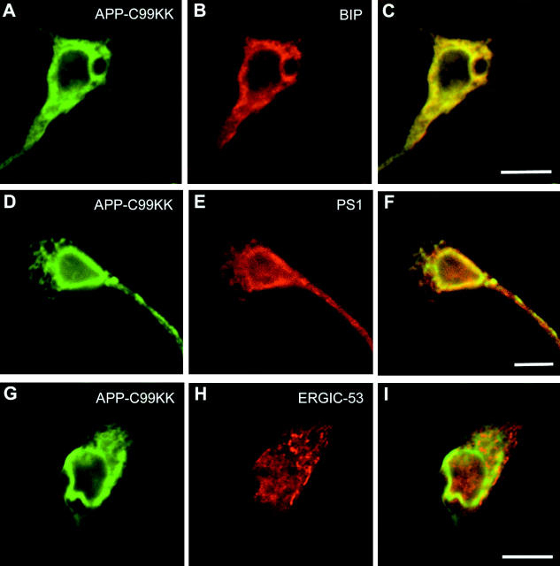
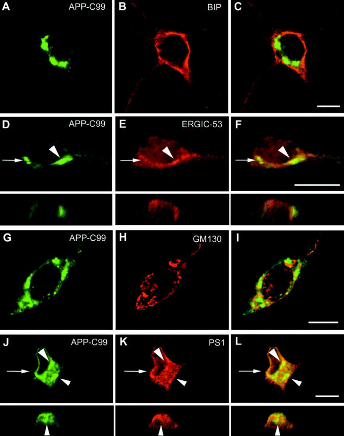
We investigated the relationship between PS1 and gamma-secretase processing of amyloid precursor protein (APP) in primary cultures of neurons. Increasing the amount of APP at the cell surface or towards endosomes did not significantly affect PS1-dependent gamma-secretase cleavage, although little PS1 is present in those subcellular compartments. In contrast, almost no gamma-secretase processing was observed when holo-APP or APP-C99, a direct substrate for gamma-secretase, were specifically retained in the endoplasmic reticulum (ER) by a double lysine retention motif. Nevertheless, APP-C99-dilysine (KK) colocalized with PS1 in the ER. In contrast, APP-C99 did not colocalize with PS1, but was efficiently processed by PS1-dependent gamma-secretase. APP-C99 resides in a compartment that is negative for ER, intermediate compartment, and Golgi marker proteins. We conclude that gamma-secretase cleavage of APP-C99 occurs in a specialized subcellular compartment where little or no PS1 is detected. This suggests that at least one other factor than PS1, located downstream of the ER, is required for the gamma-cleavage of APP-C99. In agreement, we found that intracellular gamma-secretase processing of APP-C99-KK both at the gamma40 and the gamma42 site could be restored partially after brefeldin A treatment. Our data confirm the "spatial paradox" and raise several questions regarding the PS1 is gamma-secretase hypothesis.


Figures

Figure 1.
Figure 1.
APP-trafficking mutants. APP is schematically represented (APP-WT). The different relevant proteolytic fragments are indicated at the top. The ectodomain is detected by pAb207, the amyloid peptide region 1–16 is detected by pAb B7, region 1–5 by mAb 3D6, region 17–24 is detected by mAb4G8, and the last 20 amino acids of the COOH-terminal cytoplasmic tail are detected by pAb B11/4 and pAb 6687. α-, β-, and γ-secretase cleavage sites are indicated. In the APP-C99 construct, a supplementary Asp and Ala residue (DA) has been added to obtain cleavage by the signal peptidase at the β-secretase site (see Lichtenthaler et al., 1999). For further details see Materials and methods.
Figure 2.
Figure 2.
Processing of APP trafficking mutants in PS1+/+ and PS1/neurons. (A) PS1+/+ or PS1−/− primary neuronal cultures were transduced with pSFV bearing APP-WT, APP-Δct, APP-KK, or APP-LDL as described, and metabolically labeled with [35S]methionine for 4 h at 37°C. Cell extracts (two top panels) and culture media (two bottom panels) were immunoprecipitated using the appropriate antibodies and analyzed on 10% or 4–12% NuPage gels. The position of the various APP fragments is indicated. Note that the APP-Δct holo-forms and COOH-terminal β-fragments migrate faster because the cytoplasmic domain is deleted, whereas the APP-LDL fragments migrates slower due to the large LDL receptor cytoplasmic domain. The COOH-terminal α-stub is not detected here because antibody B7 does not react with that fragment. (B) APP fragments from three independent experiments were quantified using PhosphorImaging and normalized to the level of expression of the APP holo-forms (De Strooper et al., 1995, 1998; Annaert et al., 1999). Data obtained in the PS1−/− neurons are compared with the data obtained in the PS1+/+ neurons. This shows the relative effect of the absence of PS1 on each APP fragment separately. Since APP-Δct is processed by β-secretase to a limited extent only, the results displayed in Fig. 3 are more significant for conclusions regarding the level of γ-secretase processing of this construct. (C) The secretion of Aβ42 was analyzed by ELISA. All results are normalized to the level secreted by PS1+/+ neurons transfected with APP-WT. Dark bars and light bars represent data obtained in PS1+/+ and PS1−/− neurons, respectively.
Figure 3.
Figure 3.
Secretion of p3 in PS1+/+ and/neurons expressing APP-Δct. Neurons were transduced and metabolically labeled as above. Cell extracts (top) and culture media (bottom) were immunoprecipitated using pAb207 (holo-APP) or mAb4G8 (αAPPs and Aβ/p3). The intermediate band running between Aβ and p3 in cells transduced with APP-WT corresponds to the truncated Aβ generated by β-secretase cleavage at the Glu11 position (Creemers et al., 2000).
Figure 4.
Figure 4.
γ-secretase processing of APP-C99 trafficking mutants in PS1+/+ and PS1/neurons. (A) Cells extracts (top) and culture media (bottom) of neurons expressing APP-WT, APP-C99, or APP-C99-KK were immunoprecipitated using antibodies B11/4 against the APP COOH-terminal domain (top), or B7 against the amyloid region. Arrows on the gels indicate the position of the various APP fragments. Notice the striking absence of Aβ generation with APP-C99-KK. The 6.5-kD stub seen with APP-C99 and APP-C99-KK is PS1-independent and probably not physiologically relevant, since it is never observed with APP-WT. (B) Aβ42 levels were measured by ELISA as in Fig. 2 B. Results are expressed relatively to those obtained for PS1+/+ neurons transfected with APP-WT. NS, nonsignificant (values below detection limit). (C) Aβ-fragments were quantified using PhosphorImaging and normalized to the level of expression of the APP holo-forms (n = 3; mean ± SEM). Data obtained in the PS1−/− neurons are compared with the data obtained in the PS1+/+ neurons. This shows the relative effect of the absence of PS1 on Aβ production from APP-WT and from APP-C99. The Aβ signals obtained with APP-C99-KK are below detection limit.
Figure 7.
Figure 7.
BFA treatment partially restores γ-secretase processing of APP-C99-KK. Neurons were transduced with SFV-APP-C99 or -APP-C99-KK and treated with or without 10 μM BFA for 4 h. Culture media and cell extracts were immunoprecipitated using antibody B7 and separated on 10% NuPage gels. Detection of secreted and intracellular Aβ was done by Western blotting using the W0-2 mAb raised against the NH2 terminus of the Aβ sequence (Ida et al., 1996). Neurons transduced with SFV-APP-C99-KK were treated with BFA as above. Cell extracts were sequentially immunoprecipitated using antibody FCA42, specific for Aβ peptides ending at residue 42 and FCA40, specific for Aβ peptides ending at residue 42 (Barelli et al., 1997), and separated on 10% NuPage gels. After blotting, Aβ peptides were revealed using the WO-2 mAb as above.
Figure 5.
Figure 5.
APP-C99-KK colocalizes with PS1 in the endoplasmic reticulum. Hippocampal neurons transduced with SFV-APP-C99-KK were fixed 6 h postinfection and stained with an antibody against APP (A with pab 6687; D and G with mab 3D6) and BIP (B), PS1 (E), or ERGIC-53 (H). C, F, and I represent the merged pictures. APP-C99-KK abundantly colocalized with the ER-marker protein BIP and with PS1. Little if any colocalization was observed with ERGIC-53, a resident protein of the intermediate compartment. Detection of primary antibodies was done with Alexa 488– and Alexa 546–conjugated goat anti–mouse or goat anti–rabbit secondary antibodies (1/1,000 diluted). Bar, 10 μm.
Figure 6.
Figure 6.
APP-C99 does not colocalize with PS1 and is essentially present in a BIP, ERGIC-53, and GM130 negative compartment. SFV-APP-C99 transduced hippocampal neurons were fixed and the subcellular localization of APP-C99 (A and G with pAb 6687; D and J with mAb 3D6) was compared with established marker proteins of the ER (BIP in B), the intermediate compartment (ERGIC-53 in E), the Golgi apparatus (GM130 in H), and finally with PS1 (K). C, F, I, and L show merged pictures. For D–F and J–L, the arrows point to the position of the corresponding vertical sections. Immunodetection was done as in Fig. 6. APP-C99 immunoreactivity was mainly concentrated in discrete spots that did not colocalize with the ER (C for BIP and arrowheads in L for PS1), and the Golgi region (merged panel I). Occasionally minor colocalization with ERGIC-53 could be observed (arrowheads in D–F). Bar, 10 μm.

References

    1. Annaert, W., and B. De Strooper. 1999. Presenilins: molecular switches between proteolysis and signal transduction. Trends Neurosci. 22:439–443. - PubMed
    1. Annaert, W.G., L. Levesque, K. Craessaerts, I. Dierinck, G. Snellings, D. Westaway, P.S. George-Hyslop, B. Cordell, P. Fraser, and B. De Strooper. 1999. Presenilin 1 controls γ-secretase processing of amyloid precursor protein in pre-golgi compartments of hippocampal neurons. J. Cell Biol. 147:277–294. - PMC - PubMed
    1. Barelli, H., A. Lebeau, J. Vizzanova, P. Delaere, N. Chevalier, C. Drouot, P. Marambaud, K. Ancolio, J.D. Buxbaum, O. Khorkova, et al. 1997. Characterization of new polyclonal antibodies specific for 40 and 42 amino acid long amyloid β peptides: their use to examine the cell biology of presenilins and the immunohistochemistry of sporadic Alzheimer's disease and cerebral amyloid angiopathy cases. Mol. Med. 3: 695–707. - PMC - PubMed
    1. Brou, C., F. Logeat, N. Gupta, C. Bessia, O. LeBail, J.R. Doedens, A. Cumano, P. Roux, R.A. Black, and A. Israel. 2000. A novel proteolytic cleavage involved in Notch signaling: the role of the disintegrin-metalloprotease TACE. Mol. Cell. 5:207–216. - PubMed
    1. Brown, M.S., J. Ye, R.B. Rawson, and J.L. Goldstein. 2000. Regulated intramembrane proteolysis: a control mechanism conserved from bacteria to humans. Cell. 100:391–398. - PubMed

Publication types

MeSH terms