. 2001 Aug 15;29(16):3335-46.
doi: 10.1093/nar/29.16.3335.

Characterization of Sp1, AP-1, CBF and KRC binding sites and minisatellite DNA as functional elements of the metastasis-associated mts1/S100A4 gene intronic enhancer

Affiliations

Characterization of Sp1, AP-1, CBF and KRC binding sites and minisatellite DNA as functional elements of the metastasis-associated mts1/S100A4 gene intronic enhancer

M A Cohn et al. Nucleic Acids Res. .

Abstract

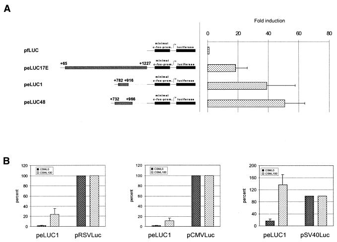
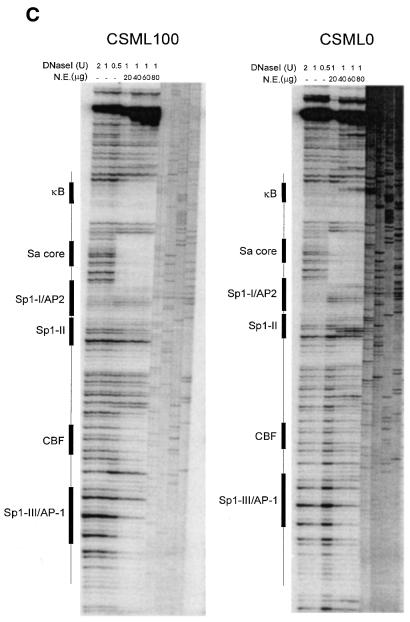
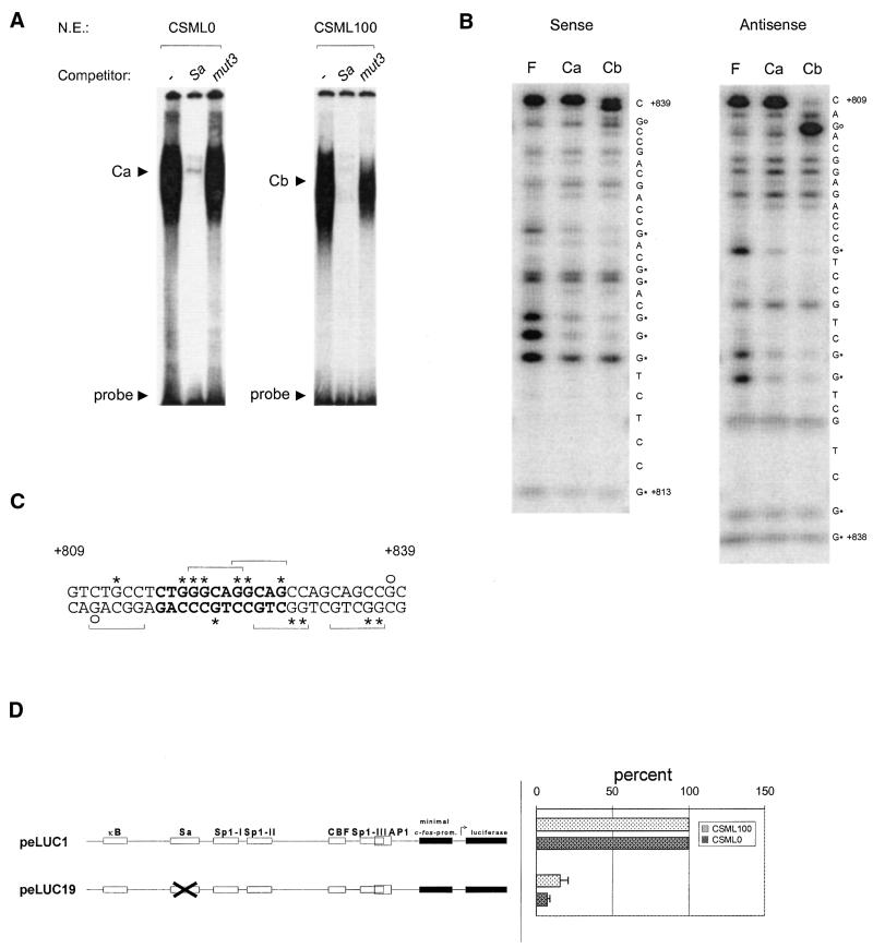
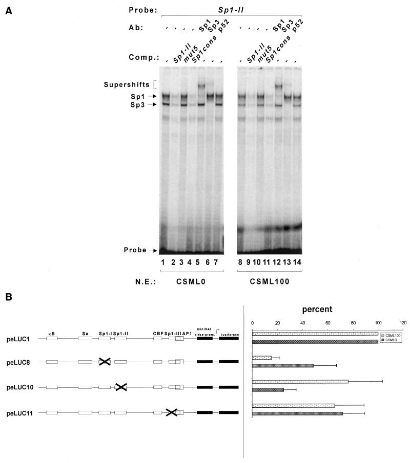
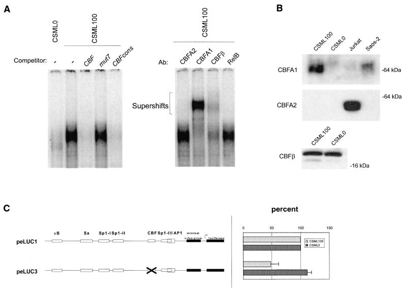
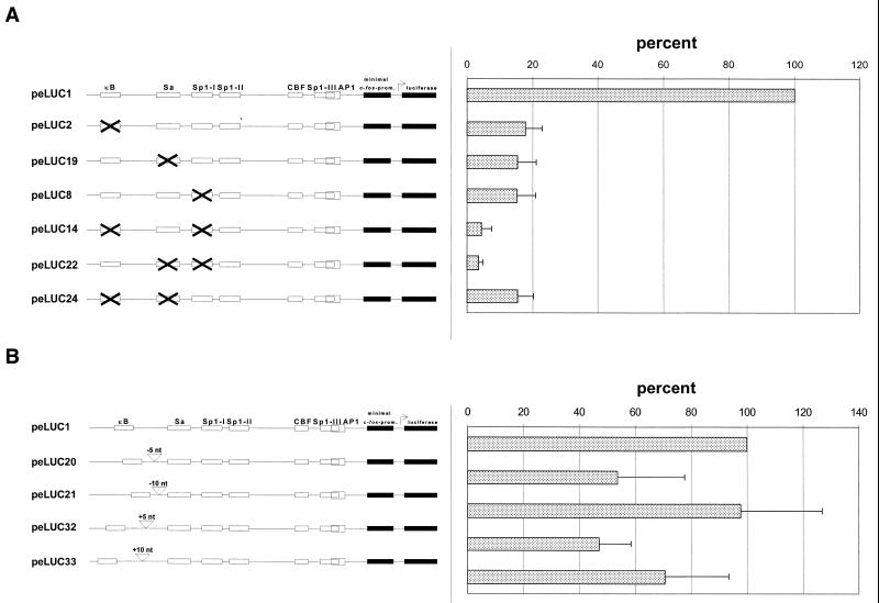
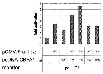
The mts1/S100A4 gene encodes a small acidic calcium-binding protein that is expressed in a cell-specific manner in development, tumorigenesis and certain tissues of adult mice. A composite enhancer that is active in murine mammary adenocarcinoma cells was previously identified in the first intron of the mts1/S100A4 gene. Here we present a detailed analysis of the structure and function of this enhancer in the Mts1/S100A4-expressing CSML100 and non-expressing CSML0 mouse adenocarcinoma cell lines. In CSML100 cells the enhancer activity is composed of at least six cis-elements interacting with Sp1 and AP-1 family members and CBF/AML/PEBP2 and KRC transcription factors. In addition, a minisatellite-like DNA sequence significantly contributes to the enhancer activity via interaction with abundant proteins, which likely have been described previously under the name minisatellite-binding proteins. Extensive mutational analysis of the mts1/S100A4 enhancer revealed a cooperative function of KRC and the factors binding minisatellite DNA. This is the first example of an enhancer where two nuclear factors earlier implicated in different recombination processes cooperate to activate transcription. In Mts1/S100A4-negative CSML0 cells the strength of the enhancer was 7- to 12.5-fold lower compared to that in CSML100 cells, when referred to the activities of three viral promoters. In CSML0 cells the enhancer could be activated by exogenous AP-1 and CBF transcription factors.


Figures

Figure 1
Figure 1
The S100A4 gene intronic enhancer is located within the 782–916 area and exhibits limited cell specificity. (A) CSML100 cells were co-transfected with reporter constructs as indicated, along with the β-galactosidase expression vector pCMVβ. Transfections were normalized for β-galactosidase activity and expressed as fold activation relative to the activity of the enhancerless pfLUC plasmid. (B) CSML0 or CSML100 cells were transfected with peLUC1 or with constructs where the luciferase gene was placed under control of the RSV (pRSVLuc), CMV (pCMVLuc) or SV40 (pSV40Luc) enhancers. Activities of the viral enhancers were taken as 100% and the activity of peLUC1 was expressed as a percentage of viral enhancer strength. Here and elsewhere the results (means ± SD) of more than three independent transfections are shown. (C) In vitro DNase I footprint analysis of the S100A4 732–966 region using CSML100 or CSML0 nuclear extracts. The non-coding strand of the DNA was labeled. The DNA was incubated with the indicated amounts of nuclear extract, followed by DNase I treatment, as shown. (D) The nucleotide sequence of the 782–916 S100A4 intronic DNA fragment. Potential transcription factor recognition sequences were identified by screening a transcription factor database (54), and are indicated by boxes. Sequences of mutant oligonucleotides used in later experiments are indicated below the sequence. The corresponding wild-type oligonucleotides used for EMSA and other experiments (Sa, Sp1-II and CBF oligonucleotides) have exactly the same length and coordinates as the corresponding mutant ones (mut3, mut5 and mut7).
Figure 1
Figure 1
The S100A4 gene intronic enhancer is located within the 782–916 area and exhibits limited cell specificity. (A) CSML100 cells were co-transfected with reporter constructs as indicated, along with the β-galactosidase expression vector pCMVβ. Transfections were normalized for β-galactosidase activity and expressed as fold activation relative to the activity of the enhancerless pfLUC plasmid. (B) CSML0 or CSML100 cells were transfected with peLUC1 or with constructs where the luciferase gene was placed under control of the RSV (pRSVLuc), CMV (pCMVLuc) or SV40 (pSV40Luc) enhancers. Activities of the viral enhancers were taken as 100% and the activity of peLUC1 was expressed as a percentage of viral enhancer strength. Here and elsewhere the results (means ± SD) of more than three independent transfections are shown. (C) In vitro DNase I footprint analysis of the S100A4 732–966 region using CSML100 or CSML0 nuclear extracts. The non-coding strand of the DNA was labeled. The DNA was incubated with the indicated amounts of nuclear extract, followed by DNase I treatment, as shown. (D) The nucleotide sequence of the 782–916 S100A4 intronic DNA fragment. Potential transcription factor recognition sequences were identified by screening a transcription factor database (54), and are indicated by boxes. Sequences of mutant oligonucleotides used in later experiments are indicated below the sequence. The corresponding wild-type oligonucleotides used for EMSA and other experiments (Sa, Sp1-II and CBF oligonucleotides) have exactly the same length and coordinates as the corresponding mutant ones (mut3, mut5 and mut7).
Figure 2
Figure 2
The minisatellite-related sequence Sa contributes to S100A4 enhancer activity. (A) EMSA showing the complexes formed by Sa in CSML0 and CSML100 nuclear extracts. A Sa-containing 31mer, the Sa oligonucleotide, was end-labeled and analyzed by EMSA with CSML0 (5 µg) and CSML100 (10 µg) nuclear extracts. Competitor oligonucleotides Sa or mut3 were added as shown. (B) Methylation interference analysis of the Ca and Cb complexes. F, free DNA. Guanines involved in formation of the Ca and Cb complexes are marked by asterisks. Circles indicate the hyper-reactive nucleotides revealed by analysis of the Cb complex. (C) Summary of the methylation interference data. The Sa core sequence is shown in bold. The 5mer motifs, GGCA/TG, are indicated by horizontal brackets. Asterisks and circles are defined in the legend to (B). (D) The contribution of Sa to the activity of the S100A4 enhancer assayed in transient transfection of CSML0 and CSML100 cells with plasmid peLUC19. peLUC19 contains a mutated Sa site (mut3). The activity of peLUC19 in CSML0 and CSML100 cells is expressed as a percentage of the wild-type enhancer activity (peLUC1) in each cell line.
Figure 3
Figure 3
Sp1-like motifs bind Sp1 and Sp3 transcription factors and differentially contribute to S100A4 enhancer function in CSML0 and CSML100 cells. (A) EMSA of a radiolabeled oligonucleotide containing the Sp1-II site with CSML0 or CSML100 nuclear extracts. Anti-Sp1, anti-Sp3 and control anti-p52 antibodies were added as indicated. Sp1 and Sp3 complexes and supershifted bands are indicated. A competition assay with unlabeled Sp1-II oligonucleotide mutant (mut5), as well as with an oligonucleotide containing a Sp1 consensus (Materials and Methods) binding site, is also shown. (B) Effect of mutating the Sp1-I, Sp1-II and Sp1-III sites on S100A4 enhancer activity. The Sp1-I, Sp1-II or Sp1-III site was substituted in peLUC1 by the mut4, mut5 or mut6 sequence (see Fig. 1D). The activity of the resulting constructs, peLUC8, peLUC10 and peLUC11, was measured in transiently transfected CSML0 or CSML100 cells and expressed as a percentage of the peLUC1 activity.
Figure 4
Figure 4
Structural and functional analysis of the CBF-binding site. (A) EMSA of complexes formed by the CBF oligonucleotide with CSML0 and CSML100 nuclear extracts. As competitors the CBF oligonucleotide, mut7 or a CBF-containing oligonucleotide derived from the Moloney murine leukemia virus enhancer (CBFcons, Materials and Methods) were used. (Right) Supershift analysis of the CBF complex formed between CBF oligonucleotide and nuclear proteins from CSML100 cells. The competitors and antibodies used are indicated above the lanes. (B) Western blot analysis of proteins from CSML0 and CSML100 cells. The antibodies used for immunostaining are indicated on the left. Nuclear extracts prepared from Saos-2 and Jurkat cells expressing CBFA1 and CBFA2 proteins, respectively, were used as positive controls. (C) Transient transfection analysis of a reporter construct with a mutated CBF site in CSML0 and CSML100 cells.
Figure 5
Figure 5
Summary of the functional analysis of the 5′-part of the S100A4 enhancer in CSML100 cells. (A) Transient transfection analysis of reporter plasmids bearing single or double point mutations in the κB, Sa and Sp1-I sites in the S100A4 enhancer. (B) Transient transfection analysis of reporter plasmids with changed phasing between the κB and Sa sites. In peLUC20 and peLUC21 5 and 10 nt are deleted, respectively. In peLUC32 and peLUC33, 5 and 10 nt are inserted, respectively.
Figure 6
Figure 6
Overexpression of the Fra-1 and CBFA1 transcription factors leads to partial reconstitution of the S100A4 enhancer in CSML0 cells. CSML0 cells were transfected with the peLUC1 reporter, along with various amounts of Fra-1 and CBFA1 expression vectors as indicated under the bars.

References

    1. Ebralidze A., Tulchinsky,E., Grigorian,M., Afanasjeva,A., Senin,V., Revazova,E. and Lukanidin,E. (1989) Isolation and characterization of a gene specifically expressed in different metastatic cells and whose deduced gene product has a high degree of homology to a Ca-binding protein family. Genes Dev., 3, 1086–1093. - PubMed
    1. Ambartsumian N.S., Grigorian,M.S., Larsen,I.F., Karlstrøm,O., Sidenius,N., Rygaard,J., Georgiev,G. and Lukanidin,E. (1996) Metastasis of mammary carcinomas in GRC/A hybrid mice transgenic for the mts1 gene. Oncogene, 13, 1621–1630. - PubMed
    1. Davies M.P.A., Rudland,P.S., Robertson,L., Parry,E.W., Jolicoeur,P. and Barraclough,R. (1996) Expression of the calcium-binding protein S100A4 (p9Ka) in MMTV-neu transgenic mice induces metastasis of mammary tumors. Oncogene, 13, 1631–1637. - PubMed
    1. Kriajevska M., Cardenas,M.N., Grigorian,M.S., Ambartsumjan,N.S., Georgiev,G. and Lukanidin,E. (1994) Non-muscle myosin heavy chain as a possible target for protein encoded by metastasis-related mts1 gene. J. Biol. Chem., 269, 19679–19682. - PubMed
    1. Bresnick A.R. (1999) Molecular mechanisms of nonmuscle myosin-II regulation. Curr. Opin. Cell Biol., 11, 26–33. - PubMed

Publication types

MeSH terms