. 2001 Aug 15;29(16):3347-55.
doi: 10.1093/nar/29.16.3347.

SOX6 binds CtBP2 to repress transcription from the Fgf-3 promoter

Affiliations

SOX6 binds CtBP2 to repress transcription from the Fgf-3 promoter

A Murakami et al. Nucleic Acids Res. .

Abstract

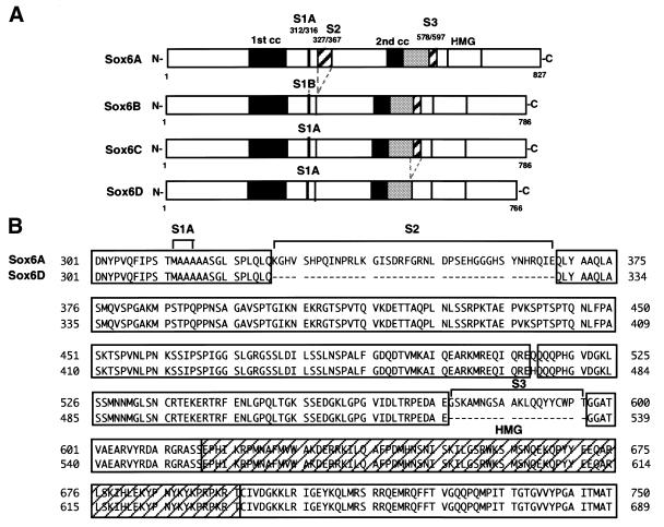
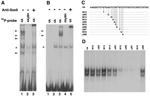
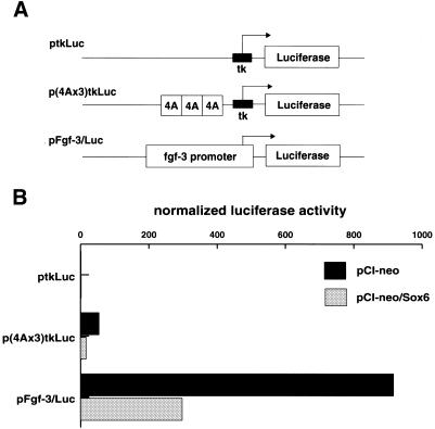
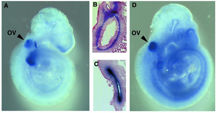
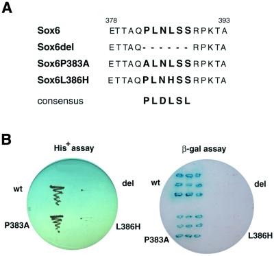
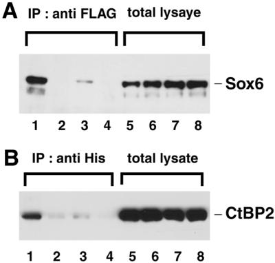
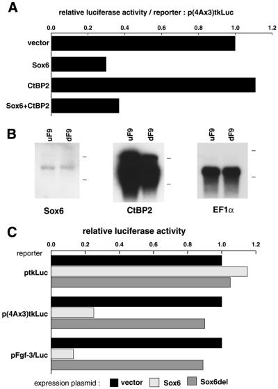
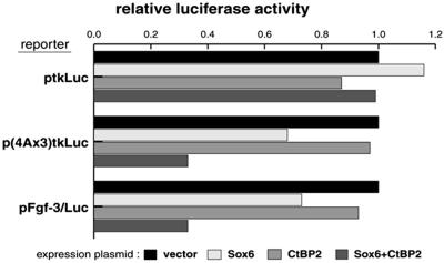
Fgf-3 is expressed in a complex pattern during mouse development. Previously, an essential regulatory element PS4A was identified in the promoter region, and shown to bind at least three factors. To identify the transcription factor(s), we used a yeast one-hybrid screen and obtained a novel Sox6 cDNA (SOX6D). When introduced into cells it strongly repressed activity from both an Fgf-3 reporter gene as well as an artificial promoter containing three PS4A elements. In situ hybridisation analysis showed that Sox6 and Fgf-3 are co-expressed in the otic vesicle of E9.5 mouse embryos in a mutually exclusive pattern, consistent with a repression of Fgf-3 transcription by SOX6. To characterise additional factor(s) involved in Fgf-3 gene repression, a yeast two-hybrid screen was used with the N-terminal portion of SOX6D. Mouse CtBP2 cDNA clones were isolated and shown to bind SOX6 in yeast and mammalian cells. Furthermore, mutational analysis of SOX6 showed that binding to CtBP2, and its responsiveness to this co-repressor, were dependent on a short amino acid sequence motif PLNLSS. Co-expression studies in NIH3T3 cells showed that SOX6 and CtBP2 co-operate to repress activity from the Fgf-3 promoter through the enhancer element PS4A. These results show that SOX6 can recruit CtBP2 to repress transcription from the Fgf-3 promoter.


Figures

Figure 1
Figure 1
Sequence comparison of SOX6 proteins. (A) Schematic comparison of SOX6 isoforms, SOX6A, SOX6B and SOX6C (28) together with the variant SOX6D. S1, S2 and S3 designate segments that differ between the isoforms. Two putative coiled-coil domains (1st cc and 2nd cc) and the HMG-box DNA binding domain common to all isoforms are indicated. (B) Comparison of the central region of SOX6A and SOX6D showing the divergent amino acid sequences. Identical residues are shaded.
Figure 2
Figure 2
Binding and recognition sequence of SOX6 proteins in PS4A. (A) EMSA using in vitro transcribed and translated SOX6 proteins and 32P-labeled probes. Lanes 1 and 3, 4A; lane 2, mutated probe 4AyM4. The + indicates addition of SOX6 antiserum after complex formation. (B) EMSA using recombinant SOX6 protein alone (lane 1) or mixed with rabbit reticulocyte lysate (lanes 3–5). Rabbit reticulocyte lysate alone (lane 2). 32P-radiolabelled probes are indicated above the lanes. Positions of complexes and a non-specific band are indicated by arrowheads and an asterisk, respectively. The two smaller complexes [lane 1 in (A)] probably arise from initiation of translation at internal AUG codons. (C) Schematic depiction showing the sequences of wild-type and point mutated PS4A probes used to establish the SOX6 binding site. (D) EMSA using 32P-labelled PS4A probes shown in (C), mixed with purified GST-Sox6DB protein.
Figure 3
Figure 3
Effects of SOX6 expression on the Fgf-3 proximal promoter and an artificial promoter containing PS4A sites. (A) Schematic representation of the control and two reporter plasmids (16) used to test the effect of SOX6 on the Fgf-3 promoter. (B) Differentiated F9 cells co-transfected with a reporter plasmid (3 µg) as indicated, and either control vector (6 µg) pCI-neo or SOX6 expression vector (6 µg) pCI-neo/Sox6. A β-galactosidase reporter (1 µg) pRL-CMV DNA was used as an internal control to normalize transfection efficiency.
Figure 4
Figure 4
Whole mount in situ hybdridisation showing Fgf-3 and Sox6 expression in mouse embryos. Mouse embryos (E9.5) were hybridised with either antisense Fgf-3 (A and B) or antisense Sox6 (C and D) probes labelled with digoxigenin and detected with anti-digoxigenin alkaline phosphatase conjugated antiserum as described in Materials and Methods. The major common expression site is in the otic vesicle (OV). Sections through the otic vesticles seen in (A) and (D) are shown in (B) and (C), respectively, and demonstrate the cellular location of the labelled cells.
Figure 5
Figure 5
Interaction of SOX6 with CtBP2 in yeast depends on the PLNLSS sequence motif in SOX6. (A) Amino acid sequences of the putative recognition site for CtBP2 in wild-type SOX6 and mutated SOX6. (B) HF7c yeast cells were co-transformed with plasmids expressing wild-type or a mutant GAL4DBD-Sox6 and GAL4AD-CtBP2. For each transformation single colonies were tested for growth on His plates (left) and for β-galactosidase activity (right), respectively.
Figure 6
Figure 6
Interaction of SOX6 with CtBP2 in Cos7 cells depends on the PLNLSS sequence in SOX6. Histidine-tagged SOX6 and FLAG-tagged CtBP2 were co-expressed in Cos7 cells. Lanes 1 and 5, SOX6 wild-type; lanes 2 and 6, SOX6del; lanes 3 and 7, SOX6P383A; lanes 4 and 8, SOX6L386H (see Fig. 5 for mutated amino acid sequences). (A) Total cell lysates (lanes 5–8) or cell lysates immunoprecipitated with anti-FLAG serum (lanes 1–4) were analysed by immunoblotting with ant-His serum. (B) As described in (A) except cell lysates were immunoprecipitated with anti-His serum and immunoblotted with anti-FLAG serum.
Figure 7
Figure 7
Effect of over-expressing CtBP2 on the repression of the Fgf-3 promoter by SOX6 in differentiated F9 cells. (A) Differentiated F9 cells were co-transfected with the p(4Ax3)tkLuc reporter together with effector plasmids for the expression of SOX6, CtBP2 or SOX6 and CtBP2. Relative luciferase activities were normalised to the value obtained with the vector alone. (B) Endogenous expression of Sox6 and CtBP2 mRNAs in F9 cells. Poly(A)+ RNA (1 µg) from undifferentiated F9 (uF9) and differentiated F9 (dF9) cells was analysed by northern blotting using 32P-labelled Sox6, CtBP2 and EF1α as a control for RNA loading. Positions of 28S and 18S ribosomal RNAs are indicated to the right of each panel. (C) SOX6 deletion mutant lacking PLNLSS does not repress transcription from the Fgf-3 promoter. Differentiated F9 cells were co-transfected with one of the reporter plasmids and an effector plasmid for expression of SOX6 or SOX6del. Relative luciferase activities are shown. Amounts of DNAs for transfection are the same as those in Figure 3.
Figure 8
Figure 8
Effects of CtBP2 on the repression by SOX6 in NIH3T3 cells. NIH3T3 cells were co-transfected with each of the reporter plasmids together with expression plasmids for the expression of SOX6 and/or CtBP2 as indicated. Relative luciferase activities are normalized to the vector alone. Amounts of DNAs for transfection are the same as those used in Figure 3.

References

    1. Ornitz D.M. (2000) FGFs, heparan sulfate and FGFRs: complex interactions essential for development. Bioessays, 22, 108–112. - PubMed
    1. McKeehan W.L., Wang,F. and Kan,M. (1998) The heparan-sulfate fibroblast growth-factor family – diversity of structure and function. Prog. Nucleic Acids Res. Mol. Biol., 59, 135–176. - PubMed
    1. Yamaguchi T.P. and Rossant,J. (1995) Fibroblast growth-factors in mammalian development. Curr. Opin. Genet. Dev., 5, 485–491. - PubMed
    1. Martin G.R. (1998) The roles of FGFs in the early development of vertebrate limbs. Genes Dev., 12, 1571–1586. - PubMed
    1. Goldfarb M. (1996) Functions of fibroblast growth factors in vertebrate development. Cytokine Growth Factor Rev., 7, 311–325. - PubMed

Publication types

MeSH terms