

doi: 10.1093/nar/29.16.e81.
DNA monolayer on gold substrates characterized by nanoparticle labeling and scanning force microscopy
Affiliations
- PMID: 11504889
- PMCID: PMC55865
- DOI: 10.1093/nar/29.16.e81
Item in Clipboard
DNA monolayer on gold substrates characterized by nanoparticle labeling and scanning force microscopy
Nucleic Acids Res.
.
Abstract
Monolayers of single-stranded DNA on gold substrates were studied by scanning force microscopy. Complementary DNA probes labeled by gold nanoparticles were applied for contrast enhancement. Substrate regions modified with DNA could be visualized in a highly specific manner. The influence of the solution concentration on the surface density of adsorbed nanoparticles could be visualized. Because individual label particles can be easily detected, this labeling technique opens the way for characterization of DNA monolayers with a lateral resolution in the nanometer range.
Figures
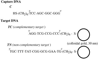
DNA sequences used in the
experiments. The thiolated capture DNA (C) was
immobilized on gold substrates. Target DNA with a sequence complementary
(TC) or non-complementary (TN)
to the capture DNA was labeled with colloidal gold (30 nm diameter).
Note that each gold colloid is covered by several DNA molecules,
which is omitted in this scheme.
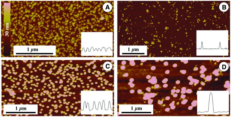
Various kinds of colloidal
gold imaged by SFM. The height is brightness coded according to
the bar in (A). Insets show cross-sections. (A)
15 nm nominal diameter; (B) Genogold 17–20
nm diameter; (C) 30 nm nominal diameter, as used
for DNA labeling, shown in Figures 3–5; (D)
60 nm nominal diameter.
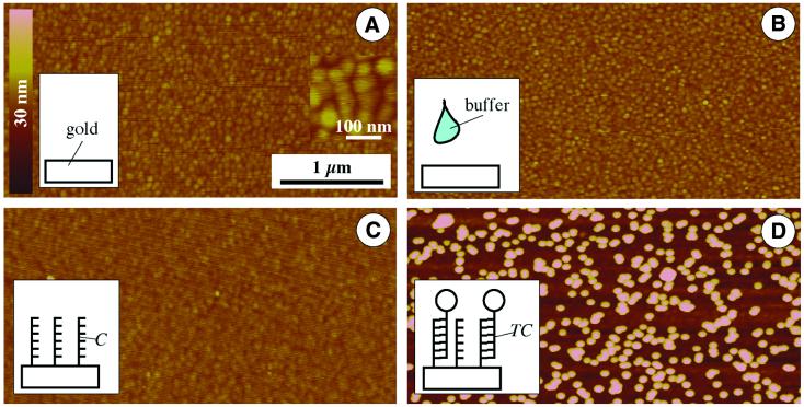
SFM imaging of the chip surface
showing different steps of substrate modification. Height and lateral
scale according to the bars in (A). (A) Pure gold substrate.
(Inset) Higher magnification revealing the grain size of the sputtered
gold. (B) Control, gold substrate incubated with
buffer solution without capture DNA. (C) Gold substrate
after incubation with capture oligonucleotides, which results in
a layer of capture probes. (D) A DNA-modified gold
substrate (as shown in C) after incubation with 30 nm gold-labeled
target probes.
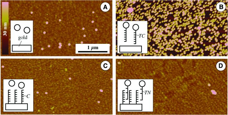
Control experiments characterizing
non-specific binding. Height and lateral scale according to the
bars in (A). (A) Pure gold substrates incubated
with pure gold colloid (without immobilized DNA). (B)
Pure gold substrate after treatment with DNA-modified gold colloid.
(C) DNA-modified surface after incubation with
pure gold solution. (D) DNA-modified substrate
treated with gold-labeled non-complementary DNA.
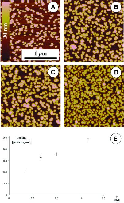
Quantification of hybridization
of gold-labeled target DNA. Solutions containing different concentrations
of labeled target DNA were incubated with surface-immobilized capture
DNA, prior to washing, air drying and SFM imaging. Concentrations
of 1 (A), 2 (B), 3 (C)
and 5 OD (D) were used. (E) Surface density
as a function of solution concentration of particles.
References
-
- Walker H.W. and Grant,S.B. (1995) Langmuir, 11, 3772–3777.
-
- Charreyre M.-T., Tcherkasskaya,O., Winnik,M.A., Hiver,A., Delair,T., Cros,P., Pichot,C. and Mandrand,B. (1997) Fluorescence energy transfer study of the conformation of oligonucleotides covalently bound to polystyrene latex particles. Langmuir, 13, 3103–3110.
-
- Peterlinz K.A., Georgiadis,R.M., Herne,T.M. and Tarlov,M.J. (1997) Observation of hybridization and dihybridization of thiol-thethered DNA using two-color surface plasmon resonance spectroscopy. J. Am. Chem. Soc., 119, 3401–3402.
-
- Herne T.M. and Tarlov,M.J. (1997) Characterization of DNA probes immobilized on gold surfaces. J. Am. Chem. Soc., 119, 8916–8920.
-
- Levicky R., Herne,T.M., Tarlov,M.J. and Satija,S.K. (1998) Using self-assembly to control the structure of DNA monolayers on gold: a neutron reflectivity study. J. Am. Chem. Soc., 120, 9787–9792.
Publication types
MeSH terms
Substances
LinkOut - more resources
Full Text Sources
Other Literature Sources
