. 2001 Aug 28;98(18):10433-8.
doi: 10.1073/pnas.181182298. Epub 2001 Aug 14.

Gene transfer of the JNK interacting protein-1 protects dopaminergic neurons in the MPTP model of Parkinson's disease

Affiliations

Gene transfer of the JNK interacting protein-1 protects dopaminergic neurons in the MPTP model of Parkinson's disease

X G Xia et al. Proc Natl Acad Sci U S A. .

Abstract

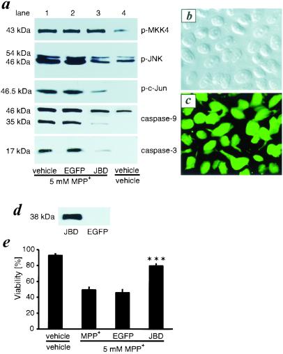
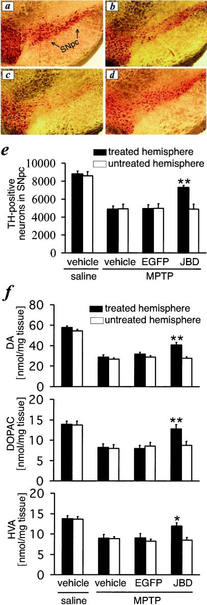
Increasing evidence suggests that apoptosis may be the underlying cell death mechanism in the selective loss of dopaminergic neurons in Parkinson's disease. Because the inhibition of caspases provides only partial protection in the 1-methyl-4-phenyl-1,2,3,6-tetrahydropyridine/1-methyl-4-phenylpyridinium (MPTP/MPP(+)) model of Parkinson's disease, we investigated the role of the proapoptotic c-Jun N-terminal kinase (JNK) signaling cascade in SH-SY5Y human neuroblastoma cells in vitro and in mice in vivo. MPTP/MPP(+) led to the sequential phosphorylation and activation of JNK kinase (MKK4), JNK, and c-Jun, the activation of caspases, and apoptosis. In mice, adenoviral gene transfer of the JNK binding domain of JNK-interacting protein-1 (a scaffold protein and inhibitor of JNK) inhibited this cascade downstream of MKK4 phosphorylation, blocked JNK, c-Jun, and caspase activation, the death of dopaminergic neurons, and the loss of catecholamines in the striatum. Furthermore, the gene transfer resulted in behavioral benefit. Therefore, inhibition of the JNK pathway offers a new treatment strategy for Parkinson's disease that blocks the death signaling pathway upstream of the execution of apoptosis in dopaminergic neurons, providing a therapeutic advantage over the direct inhibition of caspases.


Figures

Figure 1
Figure 1
JBD inhibits MPP+-induced JNK, c-Jun, and caspase activation and blocks cell death in vitro. (a) SH-SY5Y cells were infected with AdV-EGFP or AdV-JBD or were left untreated. After 48 h, these cells were treated with MPP+. Lysates of SH-SY5Y cells were collected for Western blotting 12 h later and exposed to antibodies recognizing p-MKK4, p-JNK, p-c-Jun, caspase-9, or activated caspase-3. (b) The phase contrast image was taken from AdV-JBD-infected SH-SY5Y cells. (c) Flag immunohistochemistry shows JBD-positive cells in the same culture as b. Comparing b and c, almost 100% of the cells express JBD. (d) Western blot detection of JBD expression in SH-SY5Y cells. (e) JBD overexpression rescues SH-SY5Y cells from MPP+ toxicity. ***, P < 0.001 compared with MPP+-treated or AdV-EGFP- and MPP+-treated cells (n = 6).
Figure 2
Figure 2
JBD expression inhibits MPTP-induced JNK and c-Jun but not MKK4 phosphorylation in SNpc. Detection of flag-tagged JBD expression in the striatum (a) and the SNpc (b) by immunohistochemistry or Western Blot (c) at 7 days after stereotaxic AdV-JBD injection into the striatum. Seven days after virus transfection with AdV-EGFP and AdV-JBD, mice were treated with injections of MPTP at 24-h intervals. Lysates of SNpc were collected after varying doses of MPTP and analyzed for p-MKK4, p-JNK, or p-c-Jun (d).
Figure 3
Figure 3
MPTP induces phosphorylation of c-Jun and nuclear translocation of p-c-Jun. (a) We did not detect p-c-Jun expression in SNpc dopaminergic neurons of untreated controls (green for TH, red for p-c-Jun, yellow for double staining). (b) MPTP treatment led to the induction of p-c-Jun (red) and its translocation into the nucleus (white arrow). Double staining for TH (c) revealed that the vast majority of p-c-Jun-positive cells colocalized (yellow) with dopaminergic neurons (d).
Figure 4
Figure 4
JBD blocks MPTP-induced caspase-3 activation. In TH-positive SNpc neurons (a, green) MPTP activates caspase-3 (b, red) as demonstrated by colocalization using confocal microscopy (c, yellow). JBD expression inhibited the activation of caspase-3 and the loss of TH-positive neurons (d).
Figure 5
Figure 5
JBD prevents MPTP-induced loss of TH-positive cells in SNpc and catecholamines in the striatum. Compared with controls (a), MPTP induces a loss of TH-positive neurons (b), which is not affected by prior treatment with AdV-EGFP (c). AdV-JBD prevents the loss of TH-positive cells in SNpc (d and e) and the reduction of catecholamine concentrations in the striatum (f). *, P < 0.05 and **, P < 0.01, as compared with MPTP-treated mice that previously received vehicle or AdV-EGFP injections into the striatum (n = 7–10).

References

    1. Zhang Y, Dawson V L, Dawson T M. Neurobiol Dis. 2000;7:240–250. - PubMed
    1. de Silva H R, Khan N L, Wood N W. Curr Opin Genet Dev. 2000;10:292–298. - PubMed
    1. Beal M F. Ann Neurol. 1995;38:357–366. - PubMed
    1. Jenner P, Olanow C W. Ann Neurol. 1998;44:S72–S84. - PubMed
    1. Honig L S, Rosenberg R N. Am J Med. 2000;108:317–330. - PubMed

Publication types

MeSH terms