. 2001 Aug 28;98(18):10439-44.
doi: 10.1073/pnas.181219298. Epub 2001 Aug 14.

Failed retrograde transport of NGF in a mouse model of Down's syndrome: reversal of cholinergic neurodegenerative phenotypes following NGF infusion

Affiliations

Failed retrograde transport of NGF in a mouse model of Down's syndrome: reversal of cholinergic neurodegenerative phenotypes following NGF infusion

J D Cooper et al. Proc Natl Acad Sci U S A. .

Abstract

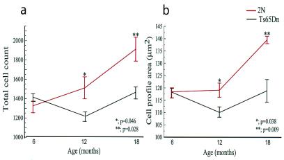
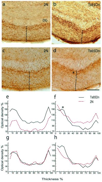
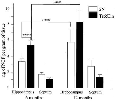
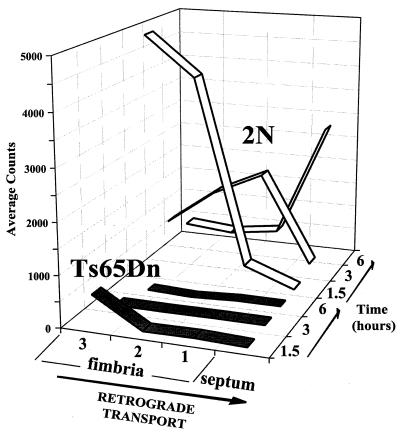
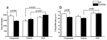
Age-related degeneration of basal forebrain cholinergic neurons (BFCNs) contributes to cognitive decline in Alzheimer's disease and Down's syndrome. With aging, the partial trisomy 16 (Ts65Dn) mouse model of Down's syndrome exhibited reductions in BFCN size and number and regressive changes in the hippocampal terminal fields of these neurons with respect to diploid controls. The changes were associated with significantly impaired retrograde transport of nerve growth factor (NGF) from the hippocampus to the basal forebrain. Intracerebroventricular NGF infusion reversed well established abnormalities in BFCN size and number and restored the deficit in cholinergic innervation. The findings are evidence that even BFCNs chronically deprived of endogenous NGF respond to an intervention that compensates for defective retrograde transport. We suggest that age-related cholinergic neurodegeneration may be a treatable disorder of failed retrograde NGF signaling.


Figures

Figure 1
Figure 1
Age-related abnormalities of BFCNs in Ts65Dn mice. (a) Plots of unbiased stereological estimates of the number of p75NGFR-immunoreactive BFCNs in the MSN (mean ± SEM) revealed no significant difference between control (2N, red line) and Ts65Dn mice (black) at 6 months of age (2N = 1328 ± 74; Ts65Dn = 1408 ± 42; P = 0.423, n = 6). In contrast, significantly fewer p75NGFR-immunoreactive BFCNs were present in Ts65Dn at 12 months of age (2N = 1512 ± 112; Ts65Dn = 1214 ± 49; P = 0.046, n = 6) and at 18 months of age (2N = 1914 ± 123; Ts65Dn = 1463 ± 63; P = 0.028, n = 5). There was a significant decrease in the number of BFCNs in Ts65Dn mice between 6 and 12 months (P = 0.028). (b) Plots of the cross-sectional area of p75NGFR-immunoreactive BFCNs (mean ± SEM) revealed no significant difference between control (2N, red line) and Ts65Dn mice (black) at 6 months of age (2N = 118.4 ± 2.5 μm2; Ts65Dn = 117.9 ± 2.0; P = 0.872, n = 6). In contrast, p75NGFR-immunoreactive BFCNs were significantly smaller in Ts65Dn mice at 12 months of age (2N = 119.13 ± 2.9 μm2; Ts65Dn = 110.2 ± 2.1; P = 0.038, n = 6) and at 18 months of age (2N = 139.5 ± 1.5 μm2; Ts65Dn = 118.8 ± 4.5; P = 0.009, n = 5). There was a significant decrease in the cell profile area in Ts65Dn BFCNs between 6 and 12 months (P = 0.016). The significance of differences between Ts65Dn and 2N mice for values in this and all subsequent figures was determined either by the Kruskal–Wallis test (multiple comparisons) or the Mann–Whitney test (paired comparisons).
Figure 2
Figure 2
Changes in the hippocampal terminal fields of BFCNs in Ts65Dn mice. Representative photomicrographs of p75NGFR-immunoreactive fibers in the molecular layer adjacent to the inferior blade of the dentate granule cell layer (DG) in 6-month-old control (2N, a) and Ts65Dn (b) mice and in 18-month-old 2N (c) and Ts65Dn (d) mice. Note the dense layer of immunoreactive fibers immediately ventral to the dentate granule cell layer at 6 months in 2N and Ts65Dn mice and at 18 months in Ts65Dn mice (*). The dotted lines denote the position of representative optical slices used to quantitatively measure p75NGFR-immunoreactive fibers. Scale bar is 50 μm. (e and f) Plots of binned optical density measurements for p75NGFR-immunoreactive fibers in the molecular layer (% thickness starts immediately adjacent to the dentate granule cell layer). The optical density in Ts65Dn (black) was higher than in 2N mice (2N, dotted red line) at 6 months and lower than 2N at 18 months. In g and h, to better compare the patterns of fiber distribution, the data shown in e and f were normalized by setting as the 100% value the optical density measurement that was registered most frequently in each group.
Figure 3
Figure 3
Septal and hippocampal NGF levels in Ts65Dn mice. ELISA measurements of NGF (nanogram of NGF per gram of tissue, wet weight; mean ± SEM) showed significant increases in the hippocampus of both 2N and Ts65Dn mice between 6 and 12 months of age. NGF levels were significantly higher in the hippocampus of Ts65Dn (black) vs. 2N (white) mice at 6 months of age (2N = 3.4 ± 0.3; Ts65Dn = 5.5 ± 0.5, P = 0.04, n = 5) but were not significantly different at 12 months of age (2N = 5.9 ± 1.6; Ts65Dn = 8.7 ± 1.9; P = 0.15, n = 4). There was a trend toward a reduction in the level of NGF in the septal region of Ts65Dn mice at 6 months of age (2N = 1.7 ± 0.2; Ts65Dn = 1.0 ± 0.2; P = 0.18, n = 5) and at 12 months of age (2N = 3.0 ± 0.2; Ts65Dn = 1.4 ± 0.3; P = 0.25, n = 4), but these changes did not reach statistical significance.
Figure 4
Figure 4
Reduced retrograde transport of 125I-NGF from hippocampus to BFCNs in Ts65Dn mice. Comparison of the retrograde transport of 125I-NGF from the hippocampus to the septum via the fimbria in 2N (white) and Ts65Dn (black) mice at 6 months of age. Radiolabeled NGF was injected into the dorsal hippocampus. Following intervals of 1.5, 3, or 6 h, the hippocampus, fimbria, and septum were dissected, and the fimbria was subdivided as defined in Materials and Methods. In 2N mice, 125I-NGF moved progressively in a wave through the proximal (i.e., closest to the hippocampus, fimbria 3), intermediate (fimbria 2), and distal segments (i.e., closest to the septum, fimbria 1) of the fimbria, arriving in the septum 6 h after injection. In contrast, in Ts65Dn mice there was little or no significant retrograde transport of 125I-NGF above background levels. The results shown are those for individual mice of each genotype at each interval for sacrifice; the results at 6 h were confirmed in two additional mice of each genotype. Failure of retrograde transport was also found in 12-month-old Ts65Dn mice (data not shown).
Figure 5
Figure 5
Restoration of BFCN number and size by NGF infusion in aged Ts65Dn mice. Comparison of stereological data for the number (a) and size (b) of p75NGFR-immunoreactive BFCNs in the MSN (mean ± SEM) of 18-month-old control (2N, white) and Ts65Dn (black) mice, either unoperated or after 2 weeks of continuous intracerebroventricular infusion of either NGF (0.9 μg/day) or artificial cerebrospinal fluid (vehicle). (a) Note that NGF treatment restored to normal the number of p75NGFR-immunoreactive BFCNs in Ts65Dn (Ts65Dn, vehicle = 1612 ± 106, n = 8; Ts65Dn, NGF = 2004 ± 119, n = 8). NGF also reversed the decrease in number that accompanied vehicle treatment of 2N mice (2N, vehicle = 1450 ± 99, n = 6; 2N, NGF = 1805 ± 76, n = 6). (b) NGF treatment also significantly increased the cross-sectional area of p75NGFR-immunoreactive BFCNs in Ts65Dn (Ts65Dn, vehicle = 104.7 ± 4.1 μm2, n = 8; Ts65Dn, NGF = 131.7 ± 4.0, n = 8). NGF also increased the profile area of BFCNs in 2N mice by 17%, to the value seen in unoperated 2N mice (2N, vehicle = 115.1 ± 5.5, n = 6; 2N, NGF = 134.6 ± 8.4, n = 6). The difference was not statistically significant.

Similar articles

Cited by

References

    1. Whitehouse P J, Price D L, Struble R G, Clark A W, Coyle J T, Delong M R. Science. 1982;215:1237–1239. - PubMed
    1. Mufson E J, Bothwell M, Kordower J H. Exp Neurol. 1989;105:221–232. - PubMed
    1. Casanova M F, Walker L C, Whitehouse P J, Price D L. Ann Neurol. 1985;18:310–313. - PubMed
    1. Mann D M, Yates P O, Marcyniuk B. Neuropathol Appl Neurobiol. 1984;10:185–207. - PubMed
    1. Reeves R H, Irving N G, Moran T H, Wohn A, Kitt C, Sisodia S S, Schmidt C, Bronson R T, Davisson M T. Nat Genet. 1995;11:177–184. - PubMed

Publication types

MeSH terms

Substances