

Comment
doi: 10.1073/pnas.181353098.
Flotillas of lipid rafts fore and aft
Affiliations
- PMID: 11504934
- PMCID: PMC55474
- DOI: 10.1073/pnas.181353098
Item in Clipboard
Comment
Flotillas of lipid rafts fore and aft
Proc Natl Acad Sci U S A.
.
No abstract available
Figures
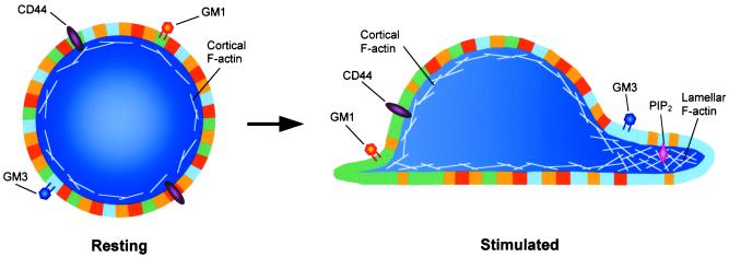
The plasma membranes of cells are composed of many types of
submicroscopic disordered (yellow regions) and more ordered (all other
regions) membrane domains, which are depicted here at much larger scale
relative to the cells than their putative size. Ordered domains are
resistant to solubilization by nonionic detergents and comprise a large
fraction of the cell surface. In resting leukocytes
(Left), all types of membrane domains, which are below
the resolution of light microscopy, are evenly distributed around the
cell periphery. Following stimulation (Right), two types
of ordered membrane domains (or rafts) segregate to either pole of the
cell, forming large assemblies (or flotillas), which can be easily
visualized by light microscopy. For ease of illustration, these
flotillas are shown as uniform patches of membrane. However, in
actuality they are more likely to be composed of ordered domains
intercalated with disordered ones. In T cells, the flotilla at the
front of the cell (blue region) is marked by the ganglioside GM3,
whereas the flotilla at the rear (green region) contains GM1. Fore and
aft flotillas may also have other compositional differences in
transmembrane proteins (e.g., CD44) and/or lipids (e.g.,
PIP2), which impart unique functions to each end of the
cell.
Comment on
-
Segregation of leading-edge and uropod components into specific lipid rafts during T cell polarization.Proc Natl Acad Sci U S A. 2001 Aug 14;98(17):9642-7. doi: 10.1073/pnas.171160298. Epub 2001 Aug 7. Proc Natl Acad Sci U S A. 2001. PMID: 11493690 Free PMC article.
References
Publication types
MeSH terms
Substances
LinkOut - more resources
Full Text Sources
