

Interchain hydrogen-bonding interactions may facilitate translocation of K+ ions across the potassium channel selectivity filter, as suggested by synthetic modeling chemistry
- PMID: 11504936
- PMCID: PMC55477
- DOI: 10.1073/pnas.161257798
Interchain hydrogen-bonding interactions may facilitate translocation of K+ ions across the potassium channel selectivity filter, as suggested by synthetic modeling chemistry
Abstract
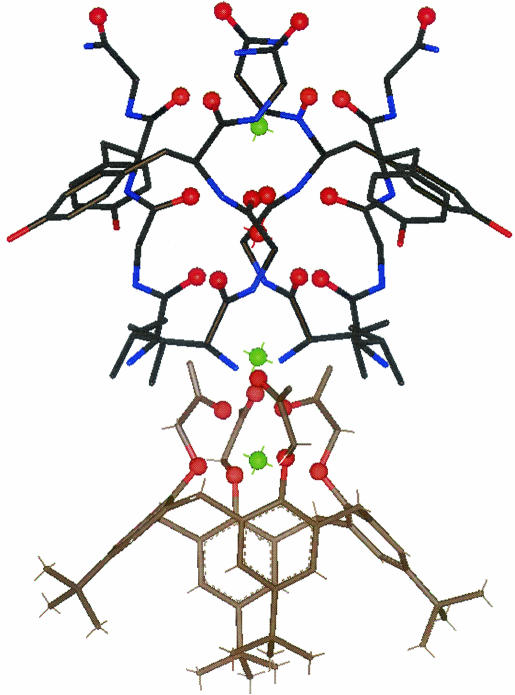
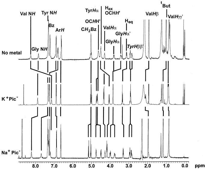
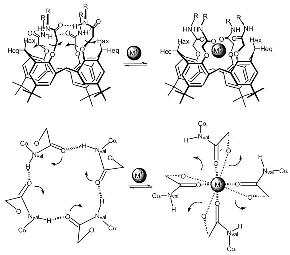
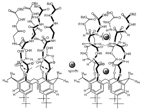
A 4-fold symmetric arrangement of TVGYG polypeptides forms the selectivity filter of the K+ channel from Streptomyces lividans (KcsA). We report the synthesis and properties of synthetic models for the filter, p-tert-butyl-calix[4]arene-(OCH(2)CO-XOBz)(4) (X = V, VG, VGY), 1-3. The first cation (Na+, K+) binds to the four -[OCH(2)CO]- units, a region devised to mimic the metal-binding site formed by the four T residues in KcsA. NMR studies reveal that cations and valine amide protons compete for the carbonyl oxygen atoms, converting NH(Val)...O=C hydrogen bonds to M+ ...O=C bonds (M+ = Na+ or K+). The strength of these interchain NH(Val)...O=C hydrogen bonds varies in the order 3 > 2 > 1. We propose that such interchain H-bonding may destabilize metal binding in the selectivity filter and thus help create the low energy barrier needed for rapid cation translocation.
Figures
References
-
- Hille B. Ionic Channels of Excitable Membranes. Sunderland, MA: Sinauer; 1992. pp. 1–19. , 115–139.
-
- Doyle D A, Cabral J M, Pfuetzner R A, Kuo A, Gulbis J M, Cohen S L, Chait B T, MacKinnon R. Science. 1998;280:69–77. - PubMed
-
- Splitt H, Meuser D, Borovok I, Betzler M, Schrempf H. FEBS Lett. 2000;472:83–87. - PubMed
Publication types
MeSH terms
Substances
LinkOut - more resources
Full Text Sources
Other Literature Sources
Medical
