Comparative Study
. 2001 Aug 14;98(17):9499-504.
doi: 10.1073/pnas.171306098.

Characterization of an adapter subunit to a phosphatidylinositol (3)P 3-phosphatase: identification of a myotubularin-related protein lacking catalytic activity

Affiliations
Comparative Study

Characterization of an adapter subunit to a phosphatidylinositol (3)P 3-phosphatase: identification of a myotubularin-related protein lacking catalytic activity

H H Nandurkar et al. Proc Natl Acad Sci U S A. .

Abstract

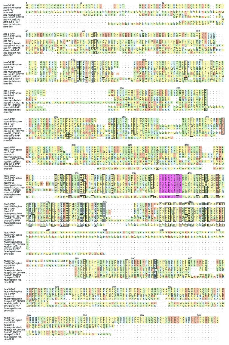
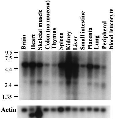
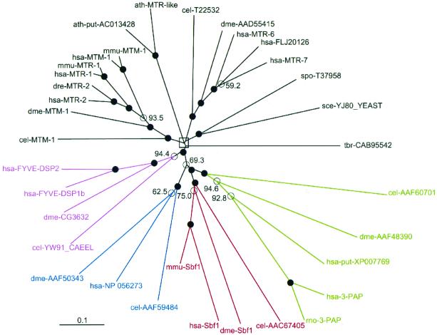
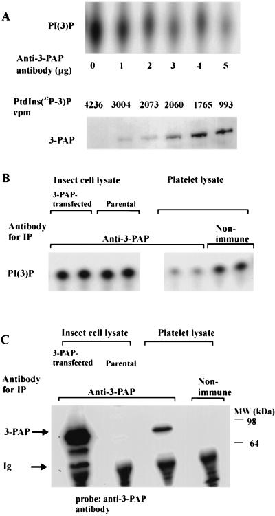
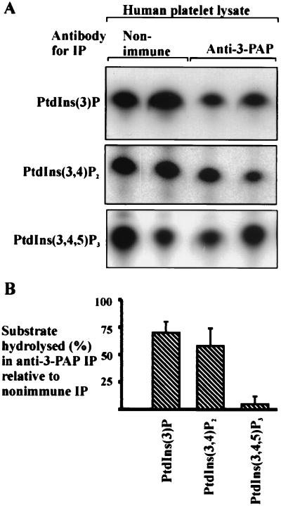
The D3-phosphoinositides act as second messengers by recruiting, and thereby activating, diverse signaling proteins. We have previously described the purification of a rat phosphatidylinositol 3-phosphate [PtdIns(3)P] 3-phosphatase, comprising a heterodimer of a 78-kDa adapter subunit in complex with a 65-kDa catalytic subunit. Here, we have cloned and characterized the cDNA encoding the human 3-phosphatase adapter subunit (3-PAP). Sequence alignment showed that 3-PAP shares significant sequence similarity with the protein and lipid 3-phosphatase myotubularin, and with several other members of the myotubularin gene family including SET-binding factor 1. However, unlike myotubularin, 3-PAP does not contain a consensus HCX(5)R catalytic motif. The 3-PAP sequence contains several motifs that predict interaction with proteins containing Src homology-2 (SH2) domains, phosphotyrosine-binding (PTB) domains, members of the 14-3-3 family, as well as proteins with SET domains. Northern blot analysis identified two transcripts (5.5 kb and 2.5 kb) with highest abundance in human liver, kidney, lung, and placenta. 3-PAP immunoprecipitates isolated from platelet cytosol hydrolyzed the D3-phosphate from PtdIns(3)P and PtdIns 3,4-bisphosphate [PtdIns(3,4)P(2)]. However, insect cell-expressed 3-PAP recombinant protein was catalytically inactive, confirming our prior prediction that this polypeptide represents an adapter subunit.


Figures

Figure 1
Figure 1
Multiple sequence alignment of the human 3-PAP with other putative homologues. Species identifiers are in italics and are as follows: hsa, Homo sapiens; rno, Rattus norvegicus; dme, D. melanogaster. Hydrophobic residues are in yellow, polar non-charged residues in green, acidic residues in brown, and basic residues in blue. Conserved residues (>70%) are boxed and in bold. The following sequences are shown: human 3-PAP, splice variant of human 3-PAP (accession no. NP 061934), rat 3-PAP, human protein similar to myotubularin-related protein-2 (accession no. XP 005992, 23% primary sequence identity); human myotubularin (accession no. XP 010178, 22% identity); human putative protein XP_007769 (38% identity); human unnamed protein NP_056273 (20% identity); Drosophila 3-PAP (Putative, accession no. AAF48390, 27% identity), human cisplatin resistance associated alpha protein (accession no. AAB36952, 23% identity), and Drosophila Sbf-1 (accession no. AAF54700, 30% identity). All sequences were identified by using PSI-blast (19), and returned highly significant Expect scores (<3 × 10−49). The active site motif (HCX5R) of myotubularin and hsa-mtr-2, and the corresponding positions in 3-PAP and related proteins are highlighted in magenta.
Figure 2
Figure 2
Tissue expression of the 3-PAP mRNA. Human multiple tissue Northern blot probed with the human expressed sequence tag R86970 corresponding to amino acids 601–747 of the composite cDNA. The migration of the RNA standards in kilobases is indicated. The blot was stripped and reexamined with an actin probe as a loading control.
Figure 3
Figure 3
Unrooted distance tree depicting the phylogenetic relationships between members of the myotubularin family. Abbreviations used are: cel, Caenorhabditis elegans; mmu, Mus musculus; dre, Danio rerio; spo, Schizosaccharomyces pombe; sce, Saccharomyces cerevisiae; tbr, Trypanosoma brucei; and ath, Arabidopsis thaliana. Protein abbreviations are as follows: MTM, myotubularin; MTR, myotubularin-related protein; FYVE-DSP, FYVE domain-containing dual specificity phosphatase; 3-PAP; 3-phosphatase adapter protein; Sbf1, SET-binding factor 1. Descriptive names are given, where defined in GenBank; otherwise GenBank accession number or SWISSPROT protein ID is shown. Nodes supported by bootstrap values at or above 95% significance threshold are indicated by filled circles; <95% are marked with open circles and the relevant values are shown. An open rectangle marks three nodes at the center of the tree for which bootstrap values were <95%; however, for the sake of clarity these values are not shown. The scale bar indicates the proportion of mutations in a sequence along a branch. The clades containing myotubularin homologues with mutations in the catalytic motif are depicted as follows: green, 3-PAP family; red, Sbf1 family; and blue, which contains the human sequence NP 056273 and related orthologues. Myotubularin homologues that contain a catalytic motif are identified in black and magenta.
Figure 4
Figure 4
3-PAP immunoprecipitates from platelets contain lipid 3-phosphatase activity. (A) Five hundred microliters of Triton X-100-soluble human platelet lysate was immunoprecipitated with 0–5 μg of anti-3-PAP antibody. The total antibody used was adjusted to 5 μg by using reciprocal quantities of nonimmune Ig, captured on protein A-Sepharose and PtdIns(3)[3-32P]P 3-phosphatase assays performed on washed immunoprecipitates. Phosphoinositides were extracted, analyzed by TLC, and measured by liquid-scintillation counting. Duplicate immunoprecipitates made with 0–5 μg of anti-3-PAP antibody were analyzed by immunoblot and probed with the anti-3-PAP antibody. (B) Triton X-100-soluble human platelet lysate (500 μl, 9 mg/ml), or Triton X-100-soluble insect Sf9 cell lysate (prepared from 12 mg of cell pellet) from either parental cells, or cells infected with 3-PAP-expressing baculovirus were incubated with 5 μg of anti-3-PAP antibody, or 5 μg nonimmune antibody as indicated. Immunoprecipitates were performed in duplicate and were analyzed for PtdIns(3)P 3-phosphatase activity. (C) Lysates prepared from Sf9 insect cells expressing recombinant 3-PAP and from parental Sf9 cells, and lysates prepared from human platelets were incubated with 5 μg of either anti-3-PAP antibody or nonimmune antibody, as indicated. Immunoblot analysis was performed by using the anti-3-PAP antibody. The migration of 3-PAP, the Ig heavy chain (Ig), and molecular weight markers (MW) (kDa) are indicated.
Figure 5
Figure 5
Immunoprecipitates of 3-PAP dephosphorylate multiple D3-phosphorylated substrates. (A) Five hundred microliters of Triton X-100-soluble platelet lysate was incubated with either 5 μg of anti-3-PAP antibody, or 5 μg nonimmune antibody and washed immunoprecipitates incubated with PtdIns(3)[3-32P]P, PtdIns(3,4)[3-32P]P2, or PtdIns(3,4,5)[3-32P]P3 in lipid phosphatase assays. (B) Immunoprecipitates were analyzed for lipid 3-phosphatase activity against the indicated phosphoinositides, as described above. Values represent mean ± SD of three experiments, each containing multiple replicates.

References

    1. Vanhaesebroeck B, Leevers S J, Panayotou G, Waterfield M D. Trends Biochem Sci. 1997;22:267–272. - PubMed
    1. Rameh L E, Cantley L C. J Biol Chem. 1999;274:8347–8350. - PubMed
    1. Czech M P. Cell. 2000;100:603–606. - PubMed
    1. Datta S R, Brunet A, Greenberg M E. Genes Dev. 1999;13:2905–2927. - PubMed
    1. Corvera S, D'Arrigo A, Stenmark H. Curr Opin Cell Biol. 1999;11:460–465. - PubMed

Publication types

MeSH terms

Associated data