

doi: 10.1073/pnas.161239298.
Propagating conformational changes over long (and short) distances in proteins
Affiliations
- PMID: 11504940
- PMCID: PMC55484
- DOI: 10.1073/pnas.161239298
Item in Clipboard
Propagating conformational changes over long (and short) distances in proteins
Proc Natl Acad Sci U S A.
.
Abstract
The problem of the propagation of conformational changes over long distances or through a closely packed protein is shown to fit a model of a ligand-induced conformational change between two protein states selected by evolution. Moreover, the kinetics of the pathway between these states is also selected so that the energy of ligand binding and the speed of the transition between conformational states are physiologically appropriate. The crystallographic data of a wild-type aspartate receptor that has negative cooperativity and a mutant that has no cooperativity but has native transmembrane signaling are shown to support this model.
Figures
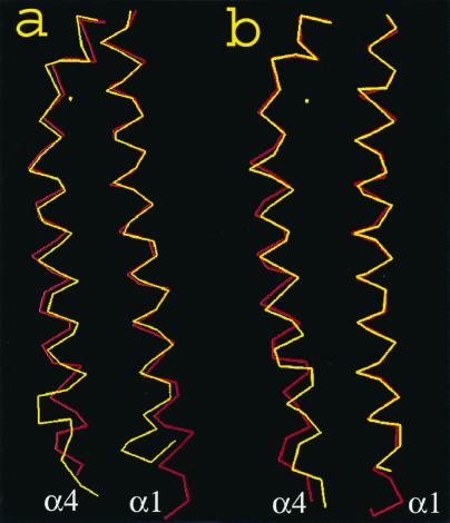
(a) The backbone atoms in the α4 helix, shown at the
left, compared with the backbone atoms of the α1 helix, shown at the
right, for the wild-type protein. The red line is the apoprotein and
the yellow line is the protein with aspartate bound. (b)
The same comparison as a, but for the S68A receptor.
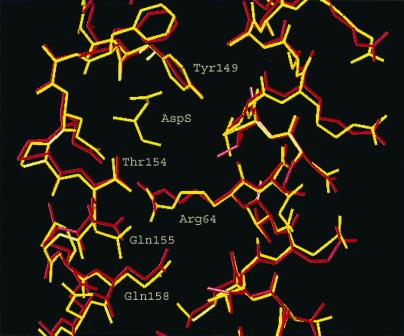
Side chains in the helices α1 and α4 that shift relative to each
other on binding aspartate.
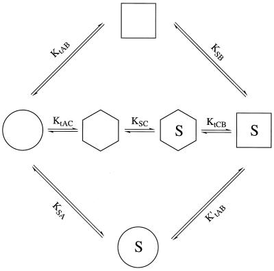
The alternative pathways for the ligand-induced change. The extremes
represent pathways in which (i) the apoprotein
isomerizes to conformation B (=
 ), which then
binds substrate
), which then
binds substrate
 (top);
(ii) the ligand binds to the apoprotein conformation that
then isomerizes to form the final bound-ligand conformation
(
(top);
(ii) the ligand binds to the apoprotein conformation that
then isomerizes to form the final bound-ligand conformation
( →
→
 →
) (bottom);
or (iii) the protein isomerizes to a conformation somewhere
in between
and
and then
binds to the ligand and reaches the final state
( →
→
) (bottom);
or (iii) the protein isomerizes to a conformation somewhere
in between
and
and then
binds to the ligand and reaches the final state
( →
 →
→
 →
)
(middle). Glossary: A =
, the
conformation of the protein in the absence of ligand. B
=
, the
conformation of the protein in the presence of ligand. C =
, an
intermediate conformation containing elements of A and B. AS =
, BS =
, CS =
, the ligand
bound to the protein in the A, B, and C conformations, respectively. Kobs = the observed affinity constant
of the receptor =
KtAB⋅KSB. KtAB = [B]/[A]. KtAC = [C]/[A]. KtCB = [B]/[C].
→
)
(middle). Glossary: A =
, the
conformation of the protein in the absence of ligand. B
=
, the
conformation of the protein in the presence of ligand. C =
, an
intermediate conformation containing elements of A and B. AS =
, BS =
, CS =
, the ligand
bound to the protein in the A, B, and C conformations, respectively. Kobs = the observed affinity constant
of the receptor =
KtAB⋅KSB. KtAB = [B]/[A]. KtAC = [C]/[A]. KtCB = [B]/[C].
), which then
binds substrate
(top);
(ii) the ligand binds to the apoprotein conformation that
then isomerizes to form the final bound-ligand conformation
( →
→
) (bottom);
or (iii) the protein isomerizes to a conformation somewhere
in between
and
and then
binds to the ligand and reaches the final state
( →
→
→
)
(middle). Glossary: A =
, the
conformation of the protein in the absence of ligand. B
=
, the
conformation of the protein in the presence of ligand. C =
, an
intermediate conformation containing elements of A and B. AS =
, BS =
, CS =
, the ligand
bound to the protein in the A, B, and C conformations, respectively. Kobs = the observed affinity constant
of the receptor =
KtAB⋅KSB. KtAB = [B]/[A]. KtAC = [C]/[A]. KtCB = [B]/[C].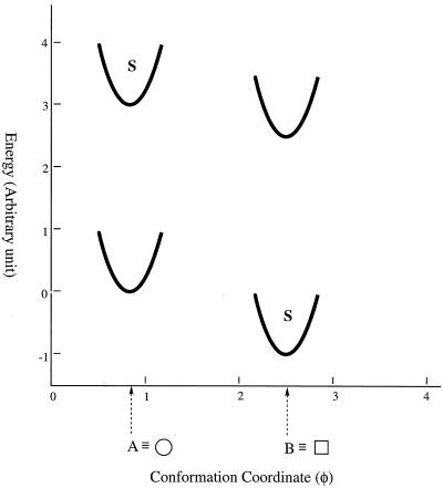
The energetics of the conformation changes when a ligand binds to a
protein. Conformational states are shown as potential wells depicting
the changing energetics of small displacement from the most stable
conformation of that well. The conformation A is the most stable
conformation of the apoprotein, but B exists at a higher energy state,
its amount depending on the kinetics and
KtAB. The presence of a ligand will
stabilize conformation B but will destabilize the A conformation
because S has very little attraction to the apo conformation.
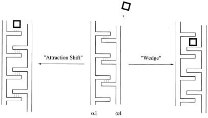
Possible mechanisms for ligand-induced changes in the aspartate
receptor. On the right is a wedge mechanism in which a ligand is
attracted into a position between rigid side chains and deflects one
helix downwards relative to the other. On the left is an attraction
shift mechanism in which the binding of a ligand attracts hydrogen
bonds, leading to a shifting of hydrogen bonds and causing the downward
shift of helix α4 relative to helix α1.
References
-
- Otteman K M, Xiao W, Shin Y-K, Koshland D E., Jr Science. 1999;285:1751–1753. - PubMed
-
- Gerstein M, Lesk A M, Chothia G. Biochemistry. 1994;33:6739–6749. - PubMed
-
- Johnson L N, Noble M E M, Owen D J. Cell. 1996;85:149–158. - PubMed
-
- Perutz M F. Nature (London) 1970;228:726–739. - PubMed
-
- Nissen P, Hansen J, Ban N, Moore P B, Steitz T A. Science. 2000;289:920–930. - PubMed
Publication types
MeSH terms
Substances
Associated data
- Actions
Grants and funding
LinkOut - more resources
Full Text Sources
Other Literature Sources
